. 2009;17(1):59-68.
doi: 10.3233/JAD-2009-1045.

Abeta-dependent Inhibition of LTP in different intracortical circuits of the visual cortex: the role of RAGE

Affiliations

Abeta-dependent Inhibition of LTP in different intracortical circuits of the visual cortex: the role of RAGE

Nicola Origlia et al. J Alzheimers Dis. 2009.

Abstract

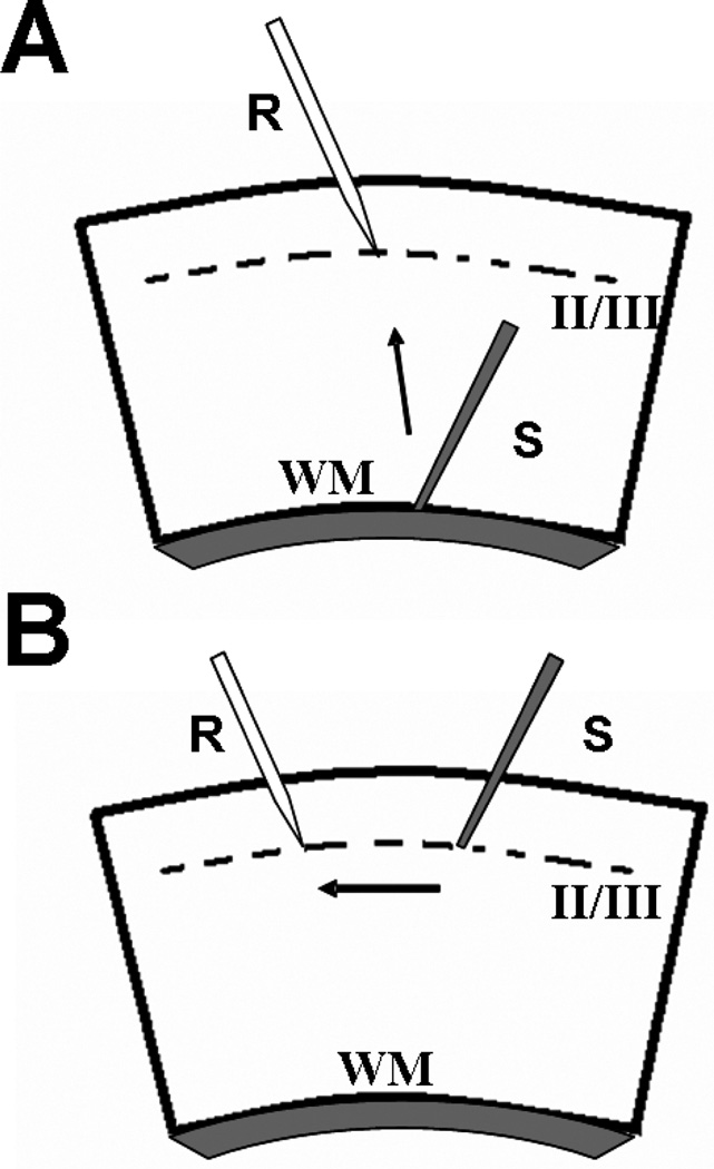
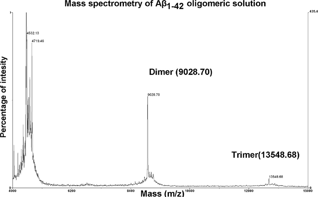
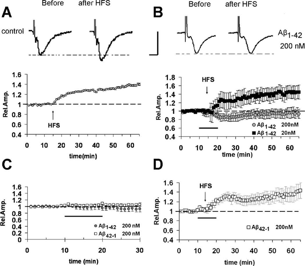
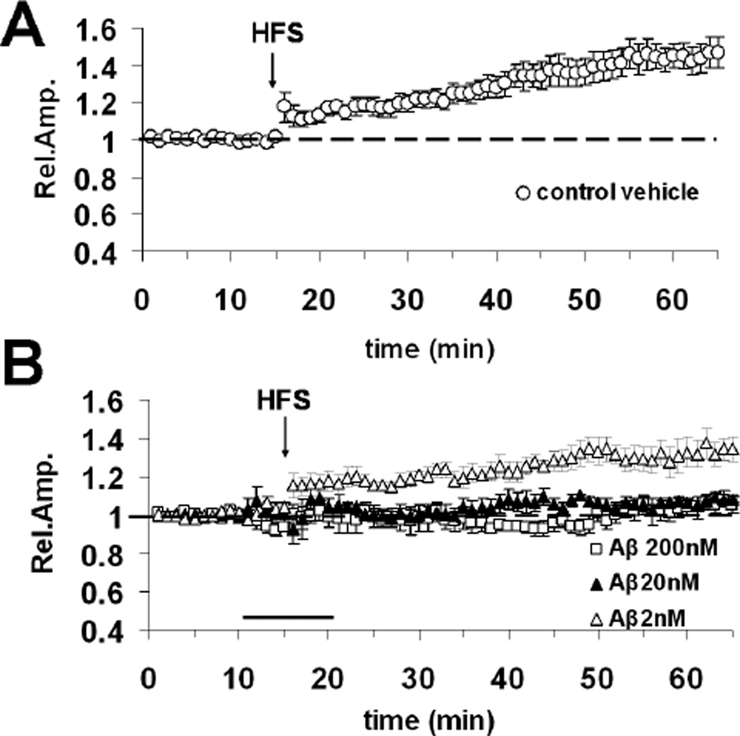
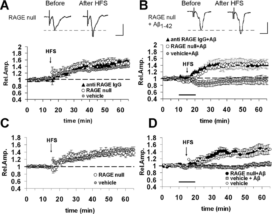
Oligomeric amyloid-beta (Abeta) interferes with long-term potentiation (LTP) and cognitive processes, suggesting that Abeta peptides may play a role in the neuronal dysfunction which characterizes the early stages of Alzheimer's disease (AD). Multiple lines of evidence have highlighted RAGE (receptor for advanced glycation end-products) as a receptor involved in Abeta-induced neuronal and synaptic dysfunction. In the present study, we investigated the effect of oligomeric soluble Abeta1-42 on LTP elicited by the stimulation of different intracortical pathways in the mouse visual cortex. A variety of nanomolar concentrations (20-200 nM) of Abeta1-42 were able to inhibit LTP in cortical layer II-III induced by either white matter (WM-Layer II/III) or the layer II/III (horizontal pathway) stimulation, whereas the inhibition of LTP was more susceptible to Abeta1-42, which occurred at 20 nM of Abeta, when stimulating layer II-III horizontal pathway. Remarkably, cortical slices were resistant to nanomolar Abeta1-42 in the absence of RAGE (genetic deletion of RAGE) or blocking RAGE by RAGE antibody. These results indicate that nanomolar Abeta inhibits LTP expression in different neocortical circuits. Crucially, it is demonstrated that Abeta-induced reduction of LTP in different cortical pathways is mediated by RAGE.


Figures

Fig. 1
Fig. 1
Schematic drawing of cortical slices and position of electrodes. (A) Representation of vertical WM-layer II/III pathway; the stimulating electrode(s) is placed at WM/layer VI boarder while the recording pipette (r) is positioned in layer II/III (corresponding to 300 µM below pial surface). (B) For stimulation of intracortical horizontal pathway, recording- (r) and stimulating- (s) electrodes were placed within the same layer (II/III).
Fig. 2
Fig. 2
Mass spectrometry analysis of Aβ1–42. Oligomeric composition of Aβ1–42 preparation was characterized by using mass spectrometry. Spectra were acquired on a Voyager-DE Pro (Applied Biosystems, Foster City, CA) as described in Materials and Methods. The main signals corresponded to the molecular ions of Aβ monomers, dimers, and trimers.
Fig. 3
Fig. 3
Inhibitory effect of Aβ1–42 on LTP elicited by the stimulation of vertical WM-Layer II/III pathway in cortical slices. (A) Under control conditions, LTP expression is induced by HFS of WM, applied after 15 minutes of baseline recording. (B) LTP is inhibited by Aβ1–42 200 nM (bath applied for 10 minutes starting from 5 minutes before HFS, dark bar; gray circles) while no effect is observed for a lower concentration of Aβ (20 nM, filled squares).(C) No effect on basal synaptic transmission was observed in the presence of Aβ1–42 (200 nM) or the reverse control peptide Aβ42–1 (200 nM) (dark bar represents application time of Aβ peptides). (D) No effect on LTP expression was observed in the presence of the control reverse peptide Aβ42–1 (200 nM). The top inserts of panels A–B show representative field potentials recorded before and 50 minutes after HFS (vertical scale bar = 0.5 mV; horizontal scale bar = 5 ms).
Fig. 4
Fig. 4
1–42 inhibits LTP elicited by the stimulation of the horizontal layer II/III ortical pathway. (A) LTP expression invehicle treated slices after HFS of layer II/III (horizontal pathway). (B) Effect of 10 minute bath application (horizontal dark bar) of different Aβ1–42 concentrations (2, 20 and 200 nM) on horizontal pathway LTP expression.
Fig. 5
Fig. 5
Blockade of RAGE protects against Aβ1–42 (200 nM)-induced impairment of cortical LTP. (A) LTP elicited by HFS of WM-Layer II/III pathway is unaffected in slices from RAGE null mice (open circles) or in slices pre-incubated with anti-RAGE IgG (2.5 µg/ml for 2 hours, filled triangles). (B) Bath application (dark bar) of Aβ42–1 (200 nM) is unable to inhibit LTP elicited by HFS of WM-Layer II/III pathway in slices from RAGE null mice (open circles) or in slices pre-incubated with anti RAGE IgG (2.5 µg/ml for 2 hours, filled triangles). In (A) and (B) top inserts show representative field potentials recorded before and 50 minutes after HFS (vertical scale bar = 0.5 mV; horiz. scale bar = 5 ms). (C) LTP elicited by HFS of horizontal layer II/III pathway is not altered in cortical slices from RAGE null mice. (D) RAGE deficiency (RAGE null slices) rescues normal LTP elicited by HFS of horizontal layer II/III pathway in Aβ-treated cortical slices (dark bar).

Similar articles

Cited by

References

    1. Gandy S. The role of cerebral amyloid beta accumulation in common forms of Alzheimer disease. J Clin Invest. 2005;115:1121–1129. - PMC - PubMed
    1. Stromer T, Serpell LC. Structure and morphology of the Alzheimer’s amyloid fibril. Microsc Res Tech. 2005;67:210–217. - PubMed
    1. Walsh DM, Klyubin I, Fadeeva JV, Cullen WK, Anwyl R, Wolfe MS, Rowan MJ, Selkoe DJ. Naturally secreted oligomers of amyloid beta protein potently inhibit hippocampal long-term potentiation in vivo . Nature. 2002;416:535–539. - PubMed
    1. Walsh DM, Townsend M, Podlisny MB, Shankar GM, Fadeeva JV, El Agnaf O, Hartley DM, Selkoe DJ. Certain inhibitors of synthetic amyloid beta-peptide (Abeta) fibrillo-genesis block oligomerization of natural Abeta and thereby rescue long-term potentiation. J Neurosci. 2005;25:2455–2462. - PMC - PubMed
    1. Wang QW, Walsh DM, Rowan MJ, Selkoe DJ, Anwyl R. Block of long-term potentiation by naturally secreted and synthetic amyloid beta-peptide in hippocampal slices is mediated via activation of the kinases c-Jun N-terminal kinase, cyclin-dependent kinase 5, and p38 mitogen-activated protein kinase as well as metabotropic glutamate receptor type 5. J Neurosci. 2004;24:3370–3378. - PMC - PubMed

Publication types

MeSH terms