. 2009;16(2):409-19.
doi: 10.3233/JAD-2009-0967.

nELAV proteins alteration in Alzheimer's disease brain: a novel putative target for amyloid-beta reverberating on AbetaPP processing

Affiliations

nELAV proteins alteration in Alzheimer's disease brain: a novel putative target for amyloid-beta reverberating on AbetaPP processing

Marialaura Amadio et al. J Alzheimers Dis. 2009.

Abstract

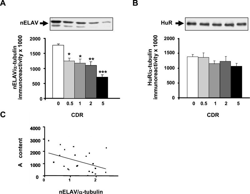
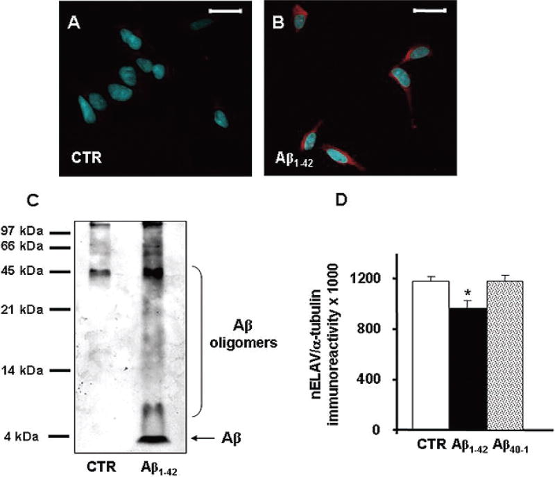
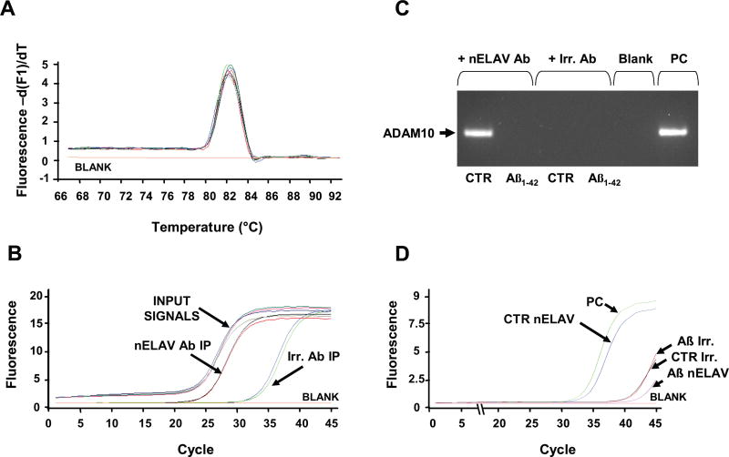
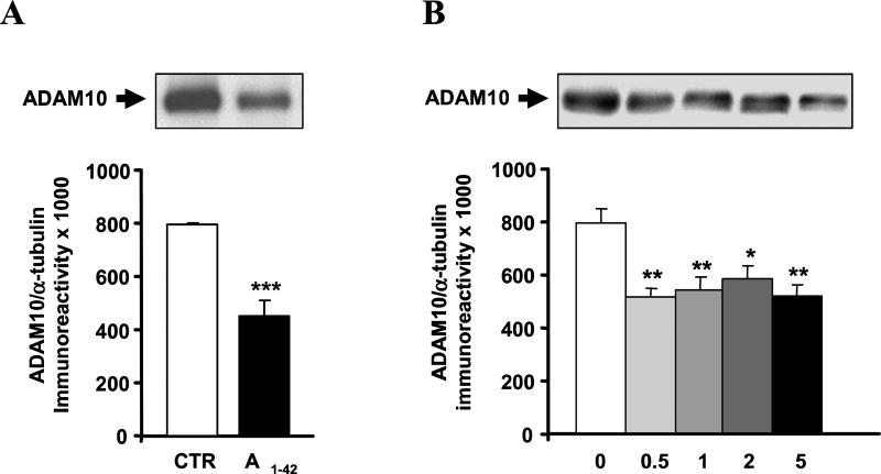
Neuronal ELAV (nELAV) proteins are RNA-binding proteins which play a physiological role in controlling gene expression in memory formation, and their alteration may contribute to cognitive impairment associated with neurodegenerative pathologies such as Alzheimer's disease (AD). Indeed, we found that the content of nELAV proteins is significantly decreased along with clinical dementia progression in the hippocampi of AD brains, where it inversely correlates with the amount of amyloid-beta (Abeta). To check the direct influence of Abeta on nELAV, we performed in vitro experiments using human SH-SY5Y cells, finding that Abeta(1-42) specifically determines nELAV proteins reduction. Since ADAM10 mRNA has the predicted sequences targeted by nELAV, we investigated whether Abeta, through nELAV proteins, could originate a vicious circle affecting amyloid-beta protein precursor (AbetaPP) processing. Immunoprecipitation experiments showed that indeed nELAV proteins bind to ADAM10 mRNA and that this binding is disrupted by Abeta(1-42) exposure, resulting in a decreased ADAM10 protein expression. ADAM10 protein diminution was also found in AD hippocampi. These data show for the first time the involvement of nELAV in AD pathology and suggest that their alteration may affect genes implicated in AbetaPP processing.


Figures

Fig. 1
Fig. 1
nELAV but not HuR protein levels are altered in the hippocampus from AD patients. (Upper) Representative Western blots of nELAV (A) and HuR (B) in the hippocampal cytoskeleton from control (CDR 0) and AD patients with a different progression of the disease (CDR 0.5, CDR 1, CDR 2, CDR 5). (Lower) Mean grey levels ratios (mean ± S.E.M.) of nELAVs/α-tubulin (A) and HuR/α-tubulin (B) immunoreactivities measured by Western blotting in the same samples. *p < 0.05, **p < 0.01, ***p < 0.001, Dunn’s test, n = 13–24. (C) nELAV proteins inversely correlate with Aβ content. Total Aβ content in the cytoskeleton of AD hippocampi was evaluated by ELISA assay. High levels of Aβ correspond to lower nELAVs/α-tubulin ratios. The solid line represents the best-fit correlation between nELAVs/α-tubulin ratios and Aβ content.
Fig. 2
Fig. 2
(A–B) Aβ is taken up by SH-SY5Y neuroblastoma cells. Confocal fluorescence representative images of SH-SY5Y control (A) and treated with 1 µMAβ1–42 (B) for 24 hours. Aβ staining (red) is almost undetectable in control cells, while in treated cells is spread all around the cytoplasm. Nuclei are stained with DAPI (blue). Scale bar: 20 µm. (C) Aβ aggregates in multiple assembly states. Representative Western blotting on the total homogenate from control and Aβ1–42 treated SH-SY5Y neuroblastoma cells. The 6E10 antibody recognizes the amino acid residues 1–17 of the Aβ sequence and is able to detect Aβ monomer and various Aβ aggregates with different molecular weights. (D) Aβ challenge reduces nELAV protein levels. (Upper) Representative Western blot of nELAV in the cytoskeleton from control and Aβ1–42 treated SH-SY5Y neuroblastoma cells. (Lower) Mean grey levels ratios (mean ± S.E.M.) of nELAV/α-tubulin immunoreactivities measured by Western blotting in the cytoskeletal fractions from the same samples. **p < 0.01, Student t-test, n = 6. (Colours are visible in the online version of the article www.iospress.nl.)
Fig. 3
Fig. 3
nELAV proteins specifically bind the ADAM10 mRNA. (A) Melting curve analysis of the Input Signals and of the immunoprecipitated pellets (IP) from nELAV and from the irrelevant antibody (Irr.). All the samples yield only one peak resulting from the specific amplification product corresponding to ADAM10. For the blank sample, the template was replaced with PCR-grade water. (B) Amplification plots showing the increase in fluorescence from nELAV IP versus Irr. IP; nELAV IP contains a much higher amount of ADAM10 template in comparison to Irr. IP [287.5 fold change calculated on the basis of the respective cycle threshold (Ct) means; nELAV IP Ct: 24.8; Irr. IP Ct: 33.2]. All the Input Signals (IS) have the same amplification plot [nELAV IS Ct: 23.6; Irr. IS Ct: 23.8], indicating that the amount of the starting template is the same for both the immunoprecipitating nELAV and Irr. antibodies. (C) Aβ challenge disrupts nELAV proteins binding to ADAM10 mRNA. Representative agarose gel following real time quantitative RT-PCR coupled with immunoprecipitation experiment using anti-nELAV antibody (+nELAV Ab) or an irrelevant antibody (+Irr. Ab), with the same isotype of nELAVs, as a negative control. The band corresponding to ADAM10 is detectable only in control SH-SY5Y cells immunoprecipitated with anti-nELAV. A cDNA obtained from a total mRNA extract was utilized as a positive control (PC). In the blank sample the template was replaced with PCR-grade water. (D) Amplification plots relative to ADAM10 contained in the samples run in Figure 3C. The plots show that nELAV Aβ1–42 treated cells IP contains a much less amount of ADAM10 template in comparison to nELAV control cells IP [respective cycle threshold (Ct): nELAV IP control, Ct: 31.7; nELAV IP Aβ1–42 treated Ct: >41; Irr. IP control Ct: >41; Irr. IP Aβ1–42 treated Ct: >41; PC Ct: 30.6].
Fig. 4
Fig. 4
(A) Aβ treatment affects ADAM10 protein levels. (Upper) Representative Western blotting of ADAM10 in the cytoskeleton from control and Aβ1–42 treated SH-SY5Yneuroblastoma cells. (Lower) Mean grey levels ratios (mean ± S.E.M.) of ADAM10/α-tubulin immunoreactivities measured by Western blotting in the cytoskeleton from the same samples. ***p < 0.001, Student t-test, n = 6. (B) ADAM10 protein levels are altered in the hippocampus from AD patients. (Upper) Representative Western blot of ADAM10 in the hippocampal cytoskeleton from control (CDR 0) and AD patients with a different progression of the disease (CDR 0.5, CDR 1, CDR 2, CDR 5). (Lower) Mean grey levels ratios (mean ± S.E.M.) of ADAM10/α-tubulin immunoreactivities measured by Western blotting in the hippocampal cytoskeleton from the same samples. *p < 0.05, **p < 0.01, Dunnett’s Multiple Comparison test, n = 13–24.

Similar articles

Cited by

References

    1. Keene JD. Why is Hu where? Shuttling of early-response-gene messenger RNA subsets. Proc Natl Acad Sci USA. 1999;96:5–7. - PMC - PubMed
    1. Pascale A, Amadio M, Quattrone A. Defining a neuron: neuronal ELAV proteins. Cell Mol Life Sci. 2008;65:128–40. - PMC - PubMed
    1. Perrone-Bizzozero N, Bolognani F. Role of HuD and other RNA-binding proteins in neural development and plasticity. J Neurosci Res. 2002;68:121–126. - PubMed
    1. Pascale A, Gusev PA, Amadio M, Dottorini T, Govoni S, Alkon DL, Quattrone A. Neuronal ELAV proteins enhance mRNA stability by a PKC alpha-dependent pathway. Proc Natl Acad Sci USA. 2004;101:1217–1222. - PubMed
    1. Quattrone A, Pascale A, Noguès X, Zhao W, Gusev P, Pacini A, Alkon DL. Posttranscriptional regulation of gene expression in learning by the neuronal ELAV-like mRNA-stabilizing proteins. Proc Natl Acad Sci USA. 2001;98:11668–11673. - PMC - PubMed

Publication types

MeSH terms