. 2009;16(2):421-31.
doi: 10.3233/JAD-2009-0980.

Cryptic peptides of the kringle domains preferentially bind to disease-associated prion protein

Affiliations

Cryptic peptides of the kringle domains preferentially bind to disease-associated prion protein

Kristen Hatcher et al. J Alzheimers Dis. 2009.

Abstract

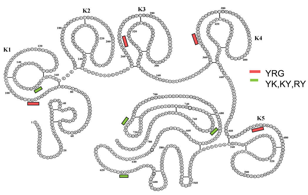
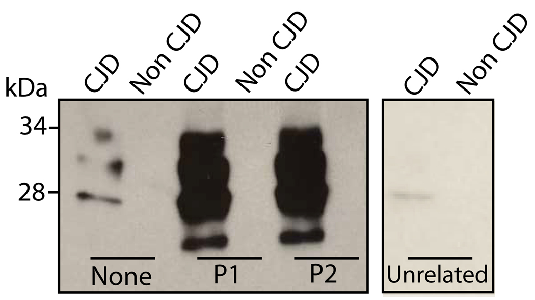
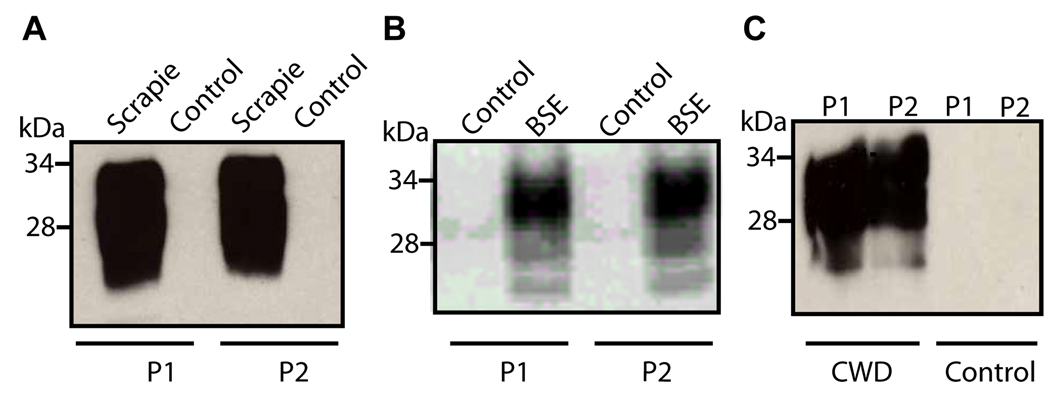
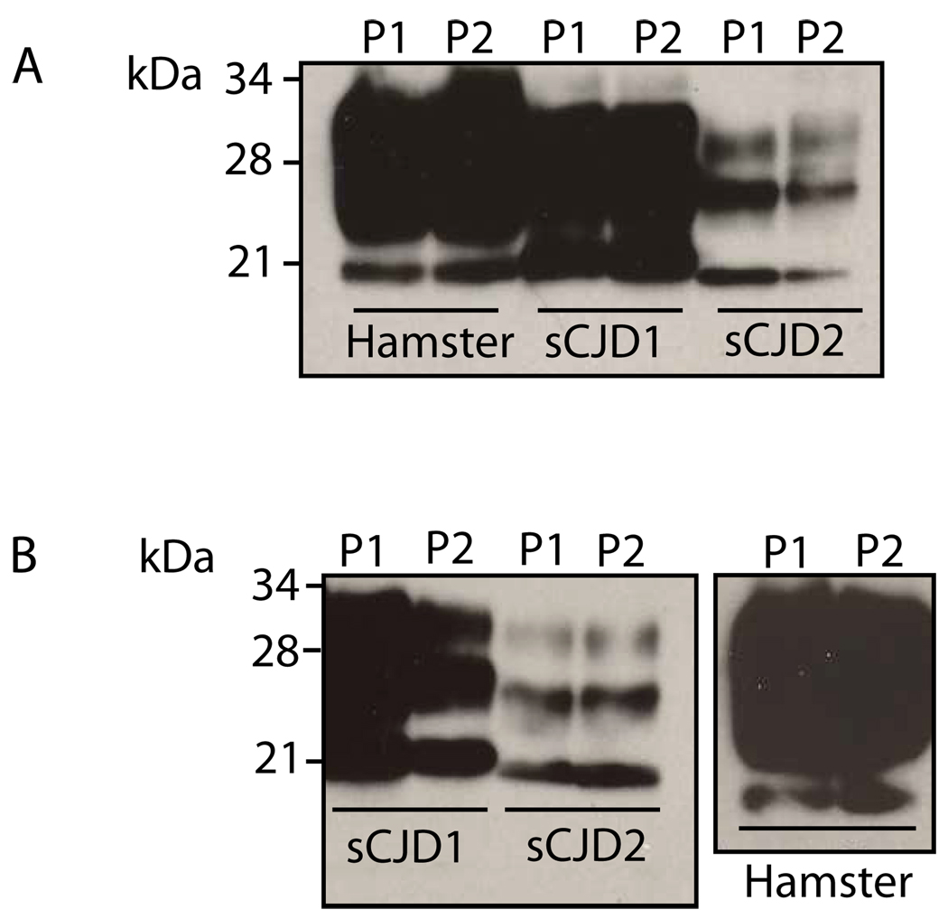
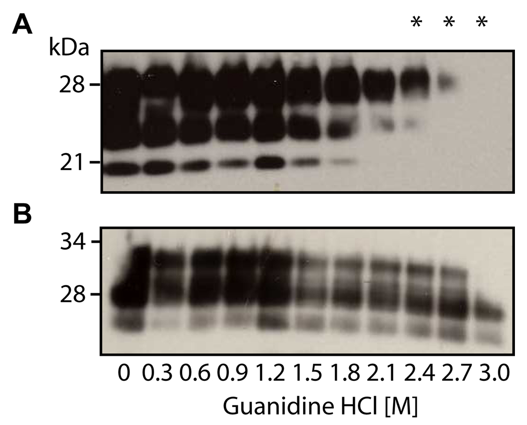
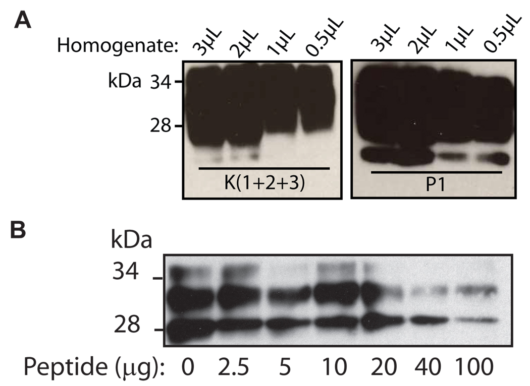
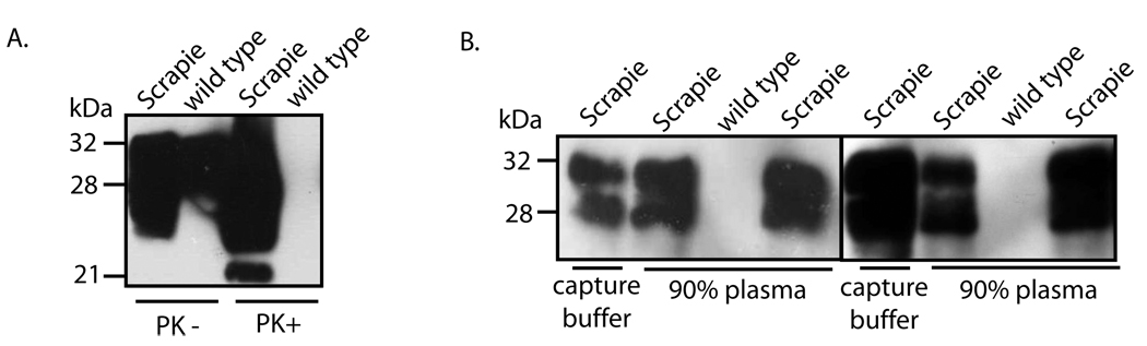
Prion diseases are a group of fatal neurodegenerative disorders characterized by the accumulation of a misfolded form (PrP(Sc)) of the cellular prion protein (PrP(C)) in the brains of affected individuals. The conversion of PrP(C) to PrP(Sc) is thought to involve a change in protein conformation from a normal, primarily alpha-helical structure into a beta-sheet conformer. Few proteins have been identified that differentially interact with the two forms of PrP. It has been reported that plasminogen binds to PrP(Sc) from a variety of prion phenotypes. We have examined potential motifs within the kringle region that may be responsible for binding to PrP. We synthesized 12-15-mer peptides that contain small, repetitive stretches of amino acid residues found within the kringle domains of plasminogen. These synthetic peptides were found to capture PrP(Sc) from the brain homogenates of bovine spongiform encephalopathy affected cattle, chronic wasting disease affected elk, experimental scrapie of hamsters and that of subjects affected by Creutzfeldt-Jakob disease, without binding to PrP(C) in unaffected controls. Therefore, we have identified critical peptide motifs that may be important for protein-protein interactions in prion disease pathogenesis. The ability of these synthetic peptides to bind preferentially to PrP(Sc) suggests a potential application in the diagnosis of prion diseases.


Figures

Fig. 1
Fig. 1. The presence of the YRG motif in the kringle domains of plasminogen
Plasminogen molecule consists of a total of five kringle domains labeled as K1–K5, and a C-terminal region. The YRG sequence appears in four out of the five kringle domains. The shorter YK, KY and RY sequences appear also in different regions of the plasminogen molecule.
Fig. 2
Fig. 2. Capture of disease-associated PrP by P1 and P2
A. P1 and P2, biotinylated and coupled to streptavidin magnetic beads, were used to capture PrP from the brain homogenates of CJD patients, as well as unaffected controls (Non-CJD). Beads with no conjugated peptide, but blocked with 0.1 % BSA, were used as a negative control (None). An unrelated peptide was biotinylated and conjugated to streptavidin magnetic beads, as a second negative control (Unrelated).
Fig. 3
Fig. 3. Capture of PrPSc from animal forms of prion disease by Peptides 1 and 2
P1 and P2, coupled to the magnetic beads, were used to pull down PrPSc in brain homogenates from Syrian hamsters infected with scrapie strain 263K (left panel), cattle affected by BSE (middle panel) and elk affected by chronic wasting disease (right panel). For the capture assay, peptide conjugated beads were incubated with 6 µl of 10 % brain homogenate in 1 ml PBS containing 3 % Tween-20 and 3 % NP-40, for three h at room temperature. Beads were recovered by applying magnetic field and proteins bound to beads were subjected to immunoblotting using either 3F4 mAb (in A) or 8H4 mAb (in B).
Fig. 4
Fig. 4. P1 and P2 capture PrPSc from different subtypes of human prion diseases
A. P1 and P2, coupled to streptavidin magnetic beads, were used to pull down PrPSc from 10% brain homogenate of individuals affected by Type 1 sCJD (Lanes 1 and 2), Type 2 sCJD (lanes 3 and 4), Type 1/2 sCJD (Lanes 5 and 6), as well as a CJD unaffected control (lanes 7 and 8), accompanied by a direct loading control of the same cases (Direct Loading). B. P1 and P2 were used to capture PrPSc from 10% brain homogenates of individuals affected by iatrogenic (lanes 1 and 2) and variant (lanes 3 and 4) CJD, as well as a CJD unaffected control (lanes 5 and 6), accompanied by a direct loading control of the same cases (Direct Loading). C. P1 and P2 were used to capture PrPSc from 10% brain homogenate from cases of familial CJD (E200K) and GSS. The 7kDa internal fragment of GSS (in C) is indicated by an asterisk. Immunoblotting was performed using 3F4 mAb.
Fig. 5
Fig. 5. Binding of P1 and P2 to the PK-resistant core fragment of PrPSc
A. Total brain homogenate from a scrapie-infected hamster (Hamster) or sporadic CJD type MM1 (sCJD1) and VV2 (sCJD2), was captured by P1 and P2 coupled to streptavidin magnetic beads, followed by digestion with 50 µg/ml PK for 1 h at 37°C and immunoblotting with 3F4. B. Total brain homogenate of sporadic CJD type MM1 (sCJD1) and VV2 (sCJD2), as well as scrapie-adapted hamster (Hamster) was digested with 50 µg ml−1 PK for 1 h at 37 °C, prior to capture by P1 and P2. Immunoblotting was performed using 3F4 mAb.
Fig. 6
Fig. 6. The binding of PrPSc to P1 is conformation dependent
A. Brain homogenate from scrapie-adapted hamsters was incubated for 1 h with guanidine HCl (0–3 M), followed by digestion with 50 µg ml−1 PK for 1 h at 37 °C and immunoblotting with 3F4.. B. Brain homogenate from scrapie-adapted hamsters was incubated for 1 h with guanidine HCl (0–3 M), followed by incubation with Peptide 1 conjugated beads and immunoblotting with 3F4. The asterisks indicate where there is a difference between PK-resistant PrP and the PrP captured directly by P1.
Fig. 7
Fig. 7. Binding of P1 to PrPSc compared to binding of angiostatin (K(1+2+3))
A. Decreasing quantities of 10 % brain homogenate from scrapie-adapted hamsters (3 µl to 0.5 µl) were incubated with 50 µg of K(1+2+3) conjugated to 2.3×10⁷ tosyl-activated beads or 7 µg of P1 conjugated 2.3×10⁷ streptavidin beads. The mixtures were incubated for 3 hours at room temperature, followed by immunoblotting with 3F4. B. Angiostatin (K(1+2+3), 1.7 mM) was conjugated to tosyl-activated magnetic beads and incubated with 4 µl of 10 % brain homogenate from a case of sporadic CJD. The binding of (K(1+2+3) to PrP was competed with increasing amounts (2–100 µg) of P1, followed by immunoblotting with 3F4.
Fig. 8
Fig. 8. Effect of substitution of arginine and tyrosine residues on the binding of P1 to PrPSc
A. The sequences of P1 and the four substituted peptides. B. The substituted peptides were conjugated to streptavidin magnetic beads and were incubated at room temperature for 3 h with 3 µl of brain homogenate from scrapie-adapted hamster, or 6 µl of brain homogenate from a normal hamster. Immunoblotting was performed using 3F4 mAb.
Fig. 9
Fig. 9. Capture of spiked PrPSc from human plasma by P1
A. Aliquots (900 µl) of human plasma obtained from two different individuals was spiked with 3 µl of 10 % total brain homogenate from hamsters infected with the 263K scrapie or from normal hamster in P1 was conjugated to streptavidin magnetic beads and were incubated at room temperature with (Lanes 1, 3, 4, and 6), followed by immunoblotting with 3F4. The conjugated beads were also incubated with 6 µl of 10 % total brain homogenate from wild type hamsters in two separate samples of human plasma (Lanes 2 and 5). Immunoblotting was performed using 3F4 mAb. B. A 10 % total brain homogenate from a hamster infected with the 263K strain of scrapie and brain homogenate from a wild type hamster were incubated in the absence (PK−) or presence (PK+) of 50 µg ml−1 PK for 1 h at 37 °C, followed by immunoblotting with 3F4.

Similar articles

Cited by

References

    1. Prusiner SB. Prions. Proc Natl Acad Sci U S A. 1998;95(23):13363–13383. - PMC - PubMed
    1. Gambetti P, et al. Sporadic and familial CJD: classification and characterisation. Br Med Bull. 2003;66:213–239. - PubMed
    1. Will RG, et al. A new variant of Creutzfeldt-Jakob disease in the UK. Lancet. 1996;347(9006):921–925. - PubMed
    1. Collen D, Lijnen HR. Basic and clinical aspects of fibrinolysis and thrombolysis. Blood. 1991;78(12):3114–3124. - PubMed
    1. Hayden SM, Seeds NW. Modulated expression of plasminogen activator system components in cultured cells from dissociated mouse dorsal root ganglia. J Neurosci. 1996;16(7):2307–2317. - PMC - PubMed

Publication types

MeSH terms