. 2009 Mar;119(3):540-50.
doi: 10.1172/JCI35541. Epub 2009 Feb 16.

Differential roles of NHERF1, NHERF2, and PDZK1 in regulating CFTR-mediated intestinal anion secretion in mice

Affiliations

Differential roles of NHERF1, NHERF2, and PDZK1 in regulating CFTR-mediated intestinal anion secretion in mice

Anurag Kumar Singh et al. J Clin Invest. 2009 Mar.

Abstract

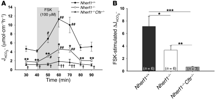
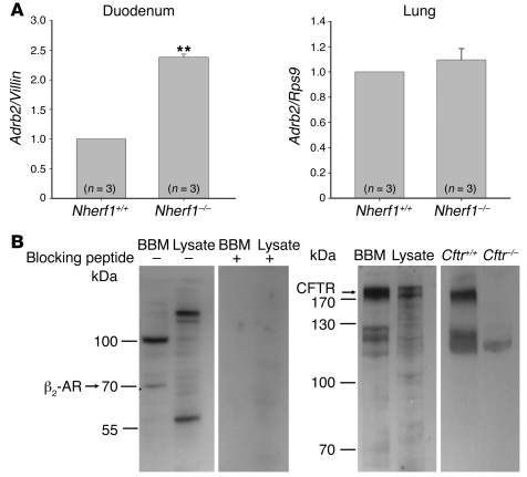
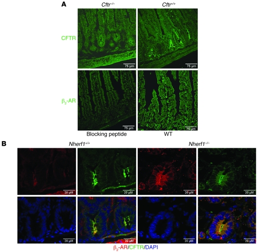
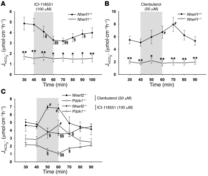
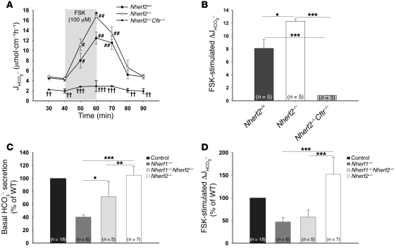
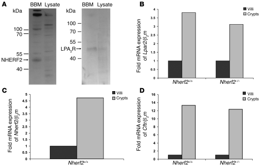
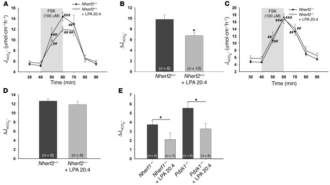
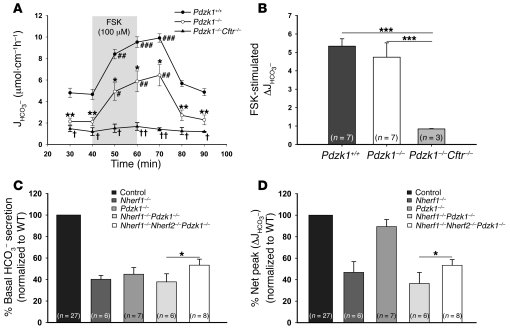
The epithelial anion channel CFTR interacts with multiple PDZ domain-containing proteins. Heterologous expression studies have demonstrated that the Na+/H+ exchanger regulatory factors, NHERF1, NHERF2, and PDZK1 (NHERF3), modulate CFTR membrane retention, conductivity, and interactions with other transporters. To study their biological roles in vivo, we investigated CFTR-dependent duodenal HCO3- secretion in mouse models of Nherf1, Nherf2, and Pdzk1 loss of function. We found that Nherf1 ablation strongly reduced basal as well as forskolin-stimulated (FSK-stimulated) HCO3- secretory rates and blocked beta2-adrenergic receptor (beta2-AR) stimulation. Conversely, Nherf2-/- mice displayed augmented FSK-stimulated HCO3- secretion. Furthermore, although lysophosphatidic acid (LPA) inhibited FSK-stimulated HCO3- secretion in WT mice, this effect was lost in Nherf2-/- mice. Pdzk1 ablation reduced basal, but not FSK-stimulated, HCO3- secretion. In addition, laser microdissection and quantitative PCR revealed that the beta2-AR and the type 2 LPA receptor were expressed together with CFTR in duodenal crypts and that colocalization of the beta2-AR and CFTR was reduced in the Nherf1-/- mice. These data suggest that the NHERF proteins differentially modulate duodenal HCO3- secretion: while NHERF1 is an obligatory linker for beta2-AR stimulation of CFTR, NHERF2 confers inhibitory signals by coupling the LPA receptor to CFTR.


Figures

Figure 1
Figure 1. Basal and FSK-stimulated HCO3 secretion in Nherf1–/–, Nherf1–/–Cftr–/–, and WT murine duodenum in vivo.
(A) Duodenal HCO3 secretion was measured in anesthetized Nherf1–/–, Nherf1–/–Cftr–/–, and WT littermates in the basal state, and during and after 20 min luminal perfusion with 10–4 M FSK. Basal as well as FSK-stimulated duodenal HCO3 secretion was dramatically reduced in the Nherf1–/– and Nherf1–/–Cftr–/– mice compared with WT littermates (n = 6 pairs of KO and WT mice, P < 0.001). The 20-min application time is denoted by shading. *P < 0.01, **P < 0.001, ***P < 0.0001 versus WT; #P < 0.01, ##P < 0.001 versus basal value; P < 0.01, ††P < 0.001 versus Nherf1–/–. (B) The net peak FSK- stimulated HCO3 secretory rate was significantly lower in Nherf1–/– and Nherf1–/–Cftr–/– mice than in WT littermates. The net peak was calculated for each experiment in this and subsequent experiments by taking the peak value and subtracting the average of the 2 basal values before the application of 10–4 M FSK. *P < 0.01; **P < 0.001; ***P < 0.0001.
Figure 2
Figure 2. Duodenal epithelial Adrb2 mRNA expression and apical membrane localization of CFTR and the β2-AR.
(A) mRNA encoding the β2-AR was expressed in murine duodenal epithelium and was significantly higher in the duodenum of Nherf1–/– than in WT mice. Conversely, no difference in Adrb2 mRNA expression level was observed in the lungs of Nherf1–/– mice. Experiments were performed in triplicate for each RNA extract from 3 pairs of mice. **P < 0.01 versus WT. (B) Western blot analysis of CFTR and the β2-AR in the BBM fraction and total cell lysate from duodenal epithelial scrapings showed that both proteins were enriched in the BBM. Both bands were blocked in the β2-AR Western blot using the blocking peptide, showing the specificity of the antibody. The higher–molecular weight band is likely a heterodimeric form of the receptor, as has been reported (62). The specificity of the CFTR band was checked using protein lysate and BBM preparation from Cftr–/– mice. The expected molecular weight of the protein is indicated.
Figure 3
Figure 3. Colocalization of CFTR and β2-AR in the apical membrane of duodenal crypts.
(A) Top: Immunohistochemical staining with an anti-CFTR antibody in Cftr–/– and Cftr+/+ duodenum showed apical crypt-predominal staining only in the Cftr+/+ tissue. Bottom: Apical staining with an antibody against β2-AR was blocked by preincubation with peptide used to raise the β2-AR antibody. β2-AR antibody stained the apical membrane of crypt as well as villous enterocytes. (B) CFTR and the β2-AR colocalized in the apical membrane of epithelial cells. In Nherf1–/– mice, the distribution of both proteins appeared more diffuse. Scale bars: 75 μm (A); 20 μm (B).
Figure 4
Figure 4. NHERF1 is essential for β2-AR–regulated duodenal HCO3 secretion.
(A and B) The luminal application of 100 μM ICI-118551 (see Methods) significantly reduced the basal HCO3 secretory rate in WT but not Nherf1–/– mice (n = 5). (B) Conversely, the luminal application of 50 μM clenbuterol (see Methods) significantly stimulated the duodenal HCO3 secretory rate in WT but not Nherf1–/– mice (n = 5). (C) Both ICI-118551 and clenbuterol had inhibitory and activating effects in Nherf2–/– and Pdzk1–/– mice, which indicates that the β2-AR–dependent regulation of CFTR is specific for NHERF1. The 20-min application time is denoted by shading. *P < 0.01, **P < 0.001 versus WT; §P < 0.01, §§P < 0.001 versus basal value (decrease); #P < 0.01 versus basal value (increase).
Figure 5
Figure 5. Basal and FSK-stimulated HCO3 secretion in Nherf2–/–, Nherf2–/–Cftr–/–, Nherf1–/–Nherf2–/–, and WT murine duodenum in vivo.
(A) Duodenal HCO3 secretion was measured as described in Figure 1. The basal HCO3 secretion in Nherf2–/– mice was unaltered, whereas the FSK-stimulated duodenal bicarbonate secretion was significantly enhanced compared with WT littermates (n = 5). Both basal and FSK-stimulated response were significantly reduced in Nherf2–/–Cftr–/– mice (n = 3). The 20-min application time is denoted by shading. *P < 0.05 versus WT; #P < 0.01, ##P < 0.001 versus basal value; P < 0.01, ††P < 0.001, †††P < 0.0001 versus Nherf2–/–. (B) The relative increase in HCO3 secretory rate was significantly higher in Nherf2–/– mice than in WT littermates (net peak, P < 0.05 and P < 0.001). (C) The absence of both Nherf1 and Nherf2 resulted in a significantly higher basal HCO3 secretory rate compared with Nherf1 deletion alone, demonstrating that NHERF2 exerts an inhibitory effect independently of NHERF1. (D) Combined Nherf1 and Nherf2 deletion neither worsened nor significantly improved the strongly reduced FSK peak secretory rate observed in Nherf1–/– mice. (BD) *P < 0.01; **P < 0.001; ***P < 0.0001.
Figure 6
Figure 6. Duodenal epithelial apical membrane localization of NHERF2 and the LPA2R and mRNA expression of Lpar2, Nherf2, and Cftr.
(A) Western blot analysis of enriched BBM and duodenal epithelial lysate using antibodies against NHERF2 and LPA2R. Both proteins were enriched in the duodenocyte BBM fraction. The expected molecular weight of the protein is indicated. (BD) The mRNA expression levels of Lpar2 (B), Nherf2 (C), and Cftr (D) were all markedly higher in the crypt region than in the villous region, indicating coexpression of these 3 proteins in the cryptal region of the duodenum. The Cftr and Lpar2 mRNA expression levels were not significantly different in the Nherf2–/– mice compared with WT.
Figure 7
Figure 7. The inhibitory effect of LPA on CFTR-mediated duodenal HCO3 secretion is absent in Nherf2–/– mice.
(AD) LPA 20:4 (50 μM), when added simultaneously with 100 μM FSK to the luminal perfusate, resulted in significant inhibition of the FSK-stimulated HCO3 secretory response in WT mice (A and B), but not in the Nherf2–/– mice (C and D), demonstrating that NHERF2 confers inhibitory signals to CFTR-mediated anion secretion in vivo. In A and C, the 20-min application time is denoted by shading. (E) In contrast, both Nherf1–/– and Pdzk1–/– mice showed a significant decrease in the FSK-stimulated duodenal bicarbonate secretion upon coperfusion with 50 μM LPA 20:4, indicating that interaction between CFTR and LPA2R is NHERF2 specific. *P < 0.05 versus respective control; #P < 0.01, ##P < 0.001, ###P < 0.001 versus basal value.
Figure 8
Figure 8. Basal bicarbonate secretion and FSK-stimulated peak secretory response in Pdzk1–/–, Pdzk1–/–Cftr–/–, Nherf1–/–Pdzk1–/–, and Nherf1–/–Nherf2–/–Pdzk1–/– duodenum.
(A and B) Basal duodenal HCO3 secretion was significantly reduced in Pdzk1–/– mice compared with WT littermates (n = 7). In A, the 20-min application time is denoted by shading. *P < 0.01, **P < 0.001 versus WT; #P < 0.01, ##P < 0.001, ###P < 0.001 versus basal value; P < 0.01, ††P < 0.001 versus Pdzk1–/–. (B) The response to FSK was not altered in Pdzk1–/– mice, but was virtually abolished in Pdzk1–/–Cftr–/– mice. (C) The additional deletion of Nherf2 resulted in a higher basal HCO3 secretory rate than in Nherf1–/– and Nherf1–/–Pdzk1–/– mice, congruent with the results in Nherf2–/– and Nherf1–/–Nherf2–/– mice. (D) The defect in FSK stimulation resulting from absence of Nherf1 was the dominant disturbance in the Nherf1–/–Pdzk1–/– and Nherf1–/–Nherf2–/–Pdzk1–/– mice. (BD) *P < 0.05; ***P < 0.001.

Similar articles

Cited by

References

    1. Guggino W.B. The cystic fibrosis transmembrane regulator forms macromolecular complexes with PDZ domain scaffold proteins. Proc. Am. Thorac. Soc. 2004;1:28–32. doi: 10.1513/pats.2306011. - DOI - PubMed
    1. Guggino W., Stanton B. New insights into cystic fibrosis: molecular switches that regulate CFTR. Nat. Rev. Mol. Cell. Biol. 2006;7:426–436. doi: 10.1038/nrm1949. - DOI - PubMed
    1. Lamprecht G., Seidler U. The emerging role of PDZ adapter proteins for regulation of intestinal ion transport. Am. J. Physiol. Gastrointest. Liver Physiol. 2006;291:G766–G777. doi: 10.1152/ajpgi.00135.2006. - DOI - PubMed
    1. Li C., Roy K., Dandridge K., Naren A.P. Molecular assembly of cystic fibrosis transmembrane conductance regulator in plasma membrane. J. Biol. Chem. 2004;279:24673–24684. doi: 10.1074/jbc.M400688200. - DOI - PubMed
    1. Moyer B.D., et al. A PDZ-interacting domain in CFTR is an apical membrane polarization signal. J. Clin. Invest. 1999;104:1353–1361. doi: 10.1172/JCI7453. - DOI - PMC - PubMed

Publication types

MeSH terms