. 2009 Mar 1;8(5):773-9.
doi: 10.4161/cc.8.5.7880. Epub 2009 Mar 18.

Logic of a mammalian metabolic cycle: an oscillated NAD+/NADH redox signaling regulates coordinated histone expression and S-phase progression

Affiliations

Logic of a mammalian metabolic cycle: an oscillated NAD+/NADH redox signaling regulates coordinated histone expression and S-phase progression

Fa-Xing Yu et al. Cell Cycle. .

Abstract

Many biological activities naturally oscillate. Here, we show that the NAD(+)/NADH ratios (redox status) fluctuate during mammalian cell cycle, with the S-phase redox status being the least oxidative. The S-phase NAD(+)/NADH redox status gates histone expression and S-phase progression, and may provide a genome protection mechanism during S-phase DNA replication as implicated in yeast. Accordingly, perturbing the cellular redox inhibits histone expression and leads to S-phase arrest. We propose that the S-phase NAD(+)/NADH redox status constitutes a redox signaling, which along with the cyclin E/cdk2 signaling regulates histone expression and S-phase progression.


Figures

Figure 1
Figure 1
Free cellular NAD+/NADH ratios fluctuate during cell cycle. (A) Random HeLa cells (R), or cells separated at the G1, S- or G2-phases were subject to FACS analyses (top); H2B mRNA levels (middle) and (free) cellular NAD+/NADH ratios (bottom) were measured. (B) The arrow underneath indicates that the re-cultured G1-phase cells progressed into the G1/S transition and S-phase (FACS analyses; top), and free cellular NAD+/NADH ratios (bottom) and H2B mRNA levels (middle) were measured.
Figure 2
Figure 2
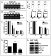
Effects of exogenous NAD(H) on H2B expression in vivo. (A–C) H2B mRNA levels in NAD(H)- treated (4 hrs) cells (A, RT-PCR [NADH]; B, RT-PCR [NAD+]; C, real-time PCR [NADH and NAD+]). (D) Free intracellular NAD+/NADH ratios effected by NAD(H) (0.5 mM, 4 hrs). (E) Cell cycle profiles in control or NAD(H)-treated cells (0.5 mM). Top, BrdU-FACS; bottom, propidium iodide (PI) staining. (F) An H2B-promoter-luciferase reporter assay in control or NAD(H)-treated cells. (G) A chromatin immunoprecipitation (ChIP) assay on control or NAD(H)-treated (0.5 mM, 4 hrs) cells to evaluate the H2B promoter occupancy by p38/GAPDH. Control IgG or anti-Oct-1, anti-TFIIB or anti-p38/GAPDH IgGs were used. The input was either 2% (NAD+) or 1% (NADH) of the starting chromatin.
Figure 3
Figure 3
An S-phase NAD+/NADH redox window. (A) H2B mRNA levels (RT-PCR) in S-phase (profile shown in Fig. 1B; top) HeLa cells untreated or treated (0.25 mM, 4 hrs) with NADH (top) or NAD+ (bottom). (B) An S-phase redox window, see text. (C) Top, free intracellular NAD+/NADH ratios in the 30–40% or 80–90% confluence HeLa cells untreated or treated with NAD(H) (4 hrs, 0.5 mM); bottom, cell cycle profiles (BrdU-FACS analyses). (D) H2B mRNA levels (real-time PCR, top; RT-PCR, bottom) in the two cell populations untreated or treated with NAD(H) (4 hrs, 0.5 mM).
Figure 4
Figure 4
A redox window in vitro or in oocytes. (A) The window (in green) is pre-established by pre-existing redox components, the removal of which allows for the biphasic response of H2B transcription to exogenously-enforced NAD+ or NAD+/NADH ratio titrations. (B) In a crude B cell nuclear extract, high exogenous NAD(H) levels disturb the pre-established redox window (see A) to inhibit the H2B transcription. The IgH template, the transcription of which is not redox-modulated,, is an internal control. (C) The H4 gene promoter is not redox-sensitive in the crude B cell nuclear extract, neither is the control (immunoglobulin kappa gene promoter).
Figure 5
Figure 5
Coordinated histone expression. (A and B) H2B and other core histone mRNA levels in control or NaN3-treated (1 mM, 2 hrs) HeLa cells as measured by RT-PCR (A) or quantified by real-time PCR (B). (C and D) correspond to (A and B) but show histone mRNA levels in control or NAD(H)-treated HeLa cells (0.5 mM, 4 hrs).
Figure 6
Figure 6
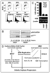
Redox status and S-phase progression. (A) Cell cycle profiles of HeLa cells treated by NAD(H) (0.5 mM, 24 hrs), analyzed by BrdU-FACS assays (top panel) and propidium iodide (PI) staining (bottom panel). (B) The p21cip1 expression levels in the redox-perturbed HeLa cells treated with NAD(H) (0.5 mM) for indicated hrs; p21cip1 levels were examined by immunoblots with p38/GAPDH as a loading control. (C) More complete H2B expression repression in the NAD(H)-treated (0.5 mM, 24 hrs) HeLa cells (bottom, RT-PCR; top, real-time PCR quantification). (D) A model emphasizing that an MMC-based redox signaling along with cyclin E/cdk2 signaling regulates histone expression and S-phase progression.

Similar articles

Cited by

References

    1. Klevecz RR, Bolen J, Forrest G, Murray DB. A genomewide oscillation in transcription gates DNA replication and cell cycle. Proc Natl Acad Sci USA. 2004;101:1200–5. - PMC - PubMed
    1. Tu BP, Kudlicki A, Rowicka M, McKnight SL. Logic of the yeast metabolic cycle: temporal compartmentalization of cellular processes. Science. 2005;310:1152–8. - PubMed
    1. Tu BP, McKnight SL. Metabolic cycles as an underlying basis of biological oscillations. Nat Rev Mol Cell Biol. 2006;9:696–701. - PubMed
    1. Xu Z, Tsurugi K. A potential mechanism of energy-metabolism oscillation in an aerobic chemostat culture of the yeast Saccharomyces cerevisiae. FEBS J. 2006;273:1696–709. - PubMed
    1. Chen Z, Odstrcil EA, Tu B, McKnight SL. Restriction of DNA replication to the reductive phase of the metabolic cycle protects genome integrity. Science. 2007;316:1916–9. - PubMed

Publication types

LinkOut - more resources