. 2009;4(2):e4473.
doi: 10.1371/journal.pone.0004473. Epub 2009 Feb 16.

Investigation of atomic level patterns in protein--small ligand interactions

Affiliations

Investigation of atomic level patterns in protein--small ligand interactions

Ke Chen et al. PLoS One. 2009.

Abstract

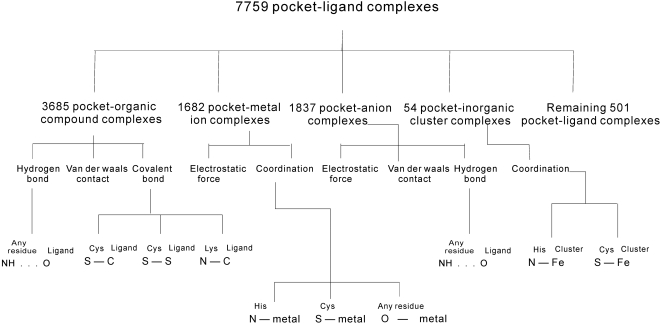
Background: Shape complementarity and non-covalent interactions are believed to drive protein-ligand interaction. To date protein-protein, protein-DNA, and protein-RNA interactions were systematically investigated, which is in contrast to interactions with small ligands. We investigate the role of covalent and non-covalent bonds in protein-small ligand interactions using a comprehensive dataset of 2,320 complexes.

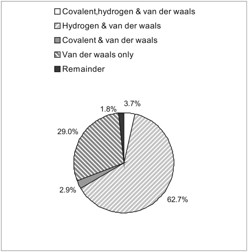
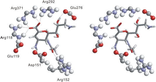
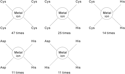
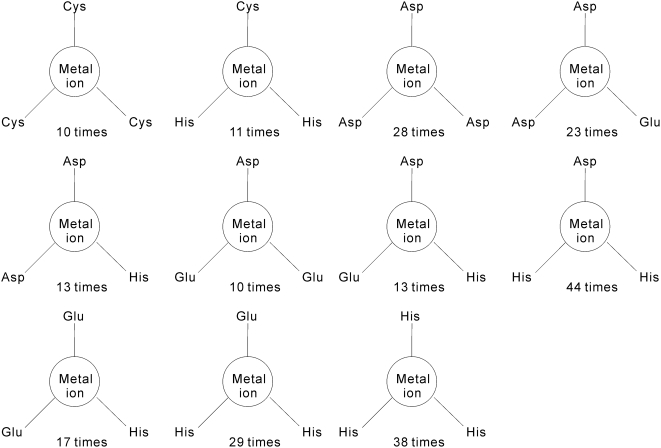
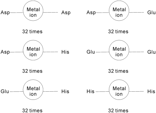
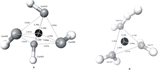
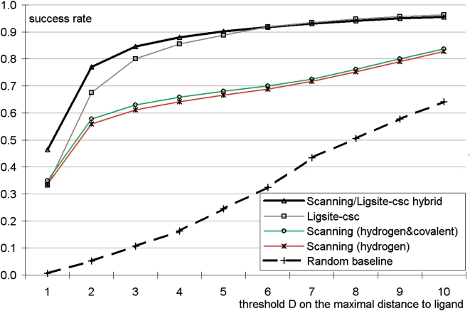
Methodology and principal findings: We show that protein-ligand interactions are governed by different forces for different ligand types, i.e., protein-organic compound interactions are governed by hydrogen bonds, van der Waals contacts, and covalent bonds; protein-metal ion interactions are dominated by electrostatic force and coordination bonds; protein-anion interactions are established with electrostatic force, hydrogen bonds, and van der Waals contacts; and protein-inorganic cluster interactions are driven by coordination bonds. We extracted several frequently occurring atomic-level patterns concerning these interactions. For instance, 73% of investigated covalent bonds were summarized with just three patterns in which bonds are formed between thiol of Cys and carbon or sulfur atoms of ligands, and nitrogen of Lys and carbon of ligands. Similar patterns were found for the coordination bonds. Hydrogen bonds occur in 67% of protein-organic compound complexes and 66% of them are formed between NH- group of protein residues and oxygen atom of ligands. We quantify relative abundance of specific interaction types and discuss their characteristic features. The extracted protein-organic compound patterns are shown to complement and improve a geometric approach for prediction of binding sites.

Conclusions and significance: We show that for a given type (group) of ligands and type of the interaction force, majority of protein-ligand interactions are repetitive and could be summarized with several simple atomic-level patterns. We summarize and analyze 10 frequently occurring interaction patterns that cover 56% of all considered complexes and we show a practical application for the patterns that concerns interactions with organic compounds.


Conflict of interest statement

Competing Interests: The authors have declared that no competing interests exist.

Figures

Figure 1
Figure 1. An overview of the protein pocket-ligand interactions.
The top layer divides protein-ligand complexes into 5 major groups based on the type of the ligand. The second layer shows the major forces that are involved in formation of protein-ligand complexes for each type of the ligand. The bottom layer summarizes significant (frequently occurring) patterns for each force/bond type and each type of the ligand. The patterns are shown in XR…YL or XR – YL format where X denotes an atom type of residue R in the protein, Y denotes an atom type of the ligand L, strong interactions (covalent and coordination bonds) are depicted by “–”, and weak interactions (hydrogen bond) are represented by “…”.
Figure 2
Figure 2. The summary of forces/bonds that are involved in formation of protein-organic compound complexes.
The chart shows that most of the complexes involve multiple contact types with the most frequent contacts involving both van der Waals force and hydrogen bonds.
Figure 3
Figure 3. An example stereo diagram of hydrogen bonds formed between NH- group of a residue and oxygen atom of an organic compound.
The oxygen atom is colored red, nitrogen atom is blue, carbon atom is gray, and hydrogen atom is white. The residues in the pocket are in ball and stick format while the ligand is in stick format. Hydrogen bonds are represented by “…”. The structure is taken from chain A of neuraminidase protein (PDB entry 1F8E), which interacts with 49A. The binding pocket contains four Arg residues and each residue contains 2 NH- groups. Three Arg residues (Arg118, Arg292, Arg371) are spatially adjacent, and they form five hydrogen bonds with the oxygen atoms of the ligand.
Figure 4
Figure 4. The residue groups that are coordinated by at least 10 metal ions and consist of 4 residues.
Figure 5
Figure 5. The residue groups that are coordinated by at least 10 metal ions and consist of 3 residues.
Figure 6
Figure 6. The residue groups that are coordinated by at least 10 metal ions and consist of 2 residues.
Figure 7
Figure 7. Examples of typical coordination bonds between metal ions and Cys and His residues.
Coordination bonds are represented by solid lines; the dashed lines show the distance between atoms of different residues. Panel A shows the coordination bond between zinc ion and four Cys residues where sulfur atom is shown in gray, carbon atom in white, and zinc ion in black. The sulfur atoms of four Cys residues form an approximate regular tetrahedron and the zinc ion is located in its center. Panel B shows the coordination bond between zinc ion and three His residues. The nitrogen atoms are shown in gray, other atoms of the His side chain are in white, and zinc ion is colored black. The three nitrogen atoms form an approximate equilateral triangle with the length of the sides that varies between 3.14 Å and 3.31 Å. The zinc ion is not located on the triangle plane.
Figure 8
Figure 8. Percentage of occurrence of amino acids in the protein-organic compound binding pockets (gray bar) and in protein-protein interaction interfaces (black bar).
Figure 9
Figure 9. Performance of blind binding site predictors including the pattern-based method, LigsiteCSC, and a random baseline predictor.
The y axis shows success rate, i.e., fraction of proteins with minimum distance between the top five predicted binding sites and any atom of a ligand in the native complex that is smaller or equal to the distance displayed on the x axis. The five plots concern the scanning method based solely on the hydrogen bond pattern (named “Scanning (hydrogen)”), the scanning method based on the four patterns concerning both hydrogen and covalent bonds (named “Scanning (hydrogen&covalent)”), the result of LigsiteCSC, the result of baseline method that randomly picks 5 solvent grid points that are within 5Å from the protein surface (named “Random baseline”), and the results that merge the top two predictions of LigsiteCSC and the top three predictions of the scanning method that uses the four patterns (named “Scanning/Ligsite-csc hybrid”).
Figure 10
Figure 10. The structure of Anguilla anguilla agglutinin protein (PDB entry 1K12).
The binding sites predicted by Ligsite CSC are colored in green and the binding sites predicted by the pattern-based method are colored in blue. The protein surface is rendered in gray and the ligand is in the stick form. The LigsiteCSC predictions are over 10Å away from any atom of the ligand, while one of pattern-based predictions is 0.67Å away from one of the ligand's atoms. Only 4 predictions by Ligsite CSC and by the pattern-based method are visible; the remaining predictions are on the other side of the protein.

References

    1. Stelzl U, Worm U, Lalowski M, Haenig C, Brembeck FH, et al. A human protein-protein interaction network: a resource for annotating the proteome. Cell. 2005;122:957–68. - PubMed
    1. Scapin G. Structural biology and drug discovery. Curr Pharm Des. 2006;12:2087–97. - PubMed
    1. Jones S, Thornton JM. Principles of protein-protein interactions. Proc Natl Acad Sci U S A. 1996;93:13–20. - PMC - PubMed
    1. Zhu H, Sommer I, Lengauer T, Domingues FS. Alignment of Non-Covalent Interactions at Protein-Protein Interfaces. PLoS ONE. 2008;3(4):e1926. - PMC - PubMed
    1. Luscombe NM, Laskowski RA, Thornton JM. Amino acid-base interactions: a three-dimensional analysis of protein-DNA interactions at an atomic level. Nucleic Acids Res. 2001;29:2860–74. - PMC - PubMed

Publication types