Review
. 2009 May;394(1):121-35.
doi: 10.1007/s00216-009-2637-8. Epub 2009 Feb 17.

Label-free technologies for quantitative multiparameter biological analysis

Affiliations
Review

Label-free technologies for quantitative multiparameter biological analysis

Abraham J Qavi et al. Anal Bioanal Chem. 2009 May.

Abstract

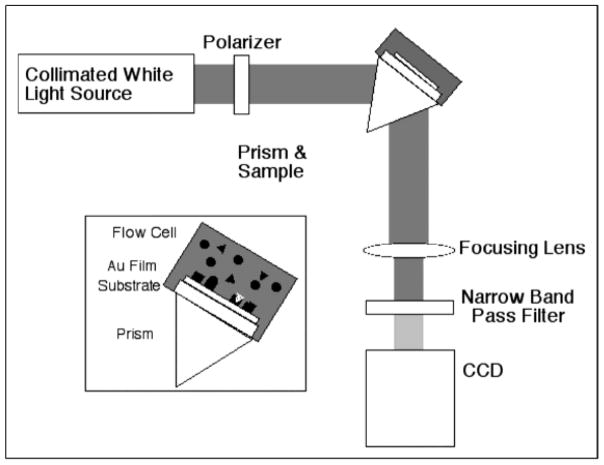
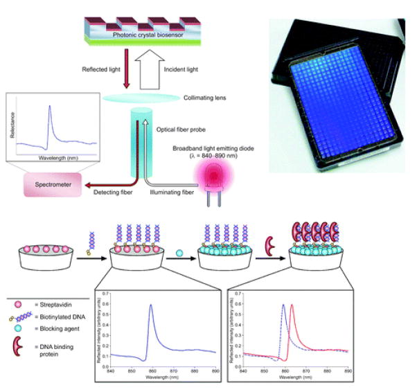
In the postgenomic era, information is king and information-rich technologies are critically important drivers in both fundamental biology and medicine. It is now known that single-parameter measurements provide only limited detail and that quantitation of multiple biomolecular signatures can more fully illuminate complex biological function. Label-free technologies have recently attracted significant interest for sensitive and quantitative multiparameter analysis of biological systems. There are several different classes of label-free sensors that are currently being developed both in academia and in industry. In this critical review, we highlight, compare, and contrast some of the more promising approaches. We describe the fundamental principles of these different methods and discuss advantages and disadvantages that might potentially help one in selecting the appropriate technology for a given bioanalytical application.


Figures

Figure 1
Figure 1
Schematic representation of an imaging surface plasmon resonance (SPRI) instrumental configuration. Biomolecular binding events are transduced as a change in reflected light intensity, and multiplexing is accomplished by imaging a large portion of the substrate using the CCD. (Figure adapted from reference [5].)
Figure 2
Figure 2
(A) SPRI image of a 120-element dsDNA array. (B) Difference image and line scan (C) after incubation of array from (A) with the transcription factor Gal4. Specific protein binding is observed as a positive change in the reflected light image. (Figure adapted from reference [16].)
Figure 3
Figure 3
(A) Schematic illustration of sample introduction onto a nanohole array biosensor. (B) Diagram showing nanohole array instrumental set up. (C) CCD image of 30 sets of nanohole arrays having different geometries. (D, E) Scanning electron micrographs showing two a top and side view of a 9 × 9 nanohole array. (Figure adapted from reference [34].)
Figure 4
Figure 4
Photonic crystal biosensors transduce biomolecular binding events by measuring the shift in wavelength of light reflected by the substrate. Shown here is a 384-well plate configuration of a photonic crystal sensing platform, which can be interrogated using a light emitting diode and simple spectrometer. This example demonstrates the screening small molecule libraries for inhibiting a specific DNA-protein binding event. (Figure adapted from reference [46].)
Figure 5
Figure 5
Photograph of a five-ringed silicon-on-insulator microring resonator array used to detect biological binding events. In this example, the microrings are accessed by on-chip waveguides that are tapered off-chip to conventional fiber optics. (Figure from reference[55].)
Figure 6
Figure 6
(A) Schematic of a Si nanowire-based FET device configured as a sensor with antibody receptors (green), where binding of a protein with net positive charge (red) yields a decrease in the conductance. (B) Cross-sectional diagram and scanning electron microscopy image of a single Si nanowire sensor device, grown via the VLS method and a photograph of a prototype nanowire sensor biochip with integrated microfluidic sample delivery. (Figure adapted from reference [85].)
Figure 7
Figure 7
A diagram (A) and scanning electron micrograph (B) of three groups of ten, 20-nm wide silicon nanowires used for label-free DNA detection. Using the superlattice nanowire patterning scheme, large numbers of precisely aligned nanowires can be fabricated for use as biosensors. (Figure from reference [99].)
Figure 8
Figure 8
Two-dimensional microcantilever array chip used to monitor protein-protein interactions. (A) Schematic of a reaction well. There were multiple cantilevers in each reaction well. Laser light reflected off a cantilever’s end pad was used to monitor the deflection of cantilevers. (B) A chip soaked in DI water. (C) A scanning electron micrograph of 3 cantilevers in a reaction well. (Figure adapted from reference [159].)
Figure 9
Figure 9
A schematic showing the principle of deflection-based microcantilever biosensing. (Figure from reference [158].)

References

    1. Kodadek T. Chemistry & Biology. 2001;8:105. - PubMed
    1. Sun YS, Landry JP, Fei YY, Zhu XD, Luo JT, Wang XB, Lam KS. Langmuir. 2008;24:13399–13405. - PMC - PubMed
    1. Sheehan PE, Whitman LJ. Nano Letters. 2005;5:803. - PubMed
    1. Squires TM, Messinger RJ, Manalis SR. Nature Biotechnology. 2008;26:417. - PubMed
    1. Smith EA, Corn RM. Applied Spectroscopy. 2003;57:320a. - PubMed

Publication types

MeSH terms

Substances

LinkOut - more resources