

Three-dimensional in vitro cell biology models of ovarian and endometrial cancer
- PMID: 19222485
- PMCID: PMC6496843
- DOI: 10.1111/j.1365-2184.2008.00579.x
Three-dimensional in vitro cell biology models of ovarian and endometrial cancer
Abstract
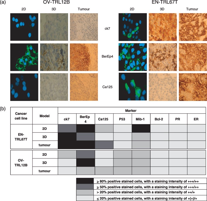
Objectives: This study aims to establish three-dimensional (3D) cell culture models of human ovarian and endometrial cancers and to compare biological and morphological characteristics of these models with those of two-dimensional (2D) models of the same cell lines and the primary tumours.
Methods: 3D models of ovarian and endometrial cancer cell cultures were established using a Rotary Cell Culture System. Immunohistochemical profiling and differential proteomics were used to characterize biological characteristics of multicellular spheroids (MCS) formed from these cultures. These were compared to characteristics of the same cells established in 2D and of the primary tumours from which the cell lines were derived.
Results: MCSs from 3D cell cultures appeared histologically similar to the primary tumours. Immunohistochemical profiling of multiple markers, including CA125, BCL2 and p53, showed that patterns of protein expression in MCSs resemble those of the primary tumours. Proteomic profiling identified several differentially expressed protein markers between 2D and 3D cultures. These included prohibitin, which was down-regulated in 3D cultures suggesting cells proliferate less compared to 2D cultures; and VDAC1 and annexin 4, which were up-regulated in 3D cultures suggesting greater levels of apoptosis in 3D compared to 2D models.
Conclusion: Establishing 3D models of cancer cell lines is likely to be of value for studying the molecular and biological mechanisms of ovarian/endometrial tumour progression and for testing novel molecular targets for cancer therapy.
Figures

 ) and EN‐TRL67T (
 ) over a period of 60 days in culture. (b) Colony‐forming efficiency (CFE%) and (c) invasive ability (cell number) of OV‐TRL12B and EN‐TRL67T cell lines. OV‐TRL12B cells showed a 3% CFE, EN‐TRL67T a CFE of 0.5%, and both cell lines also showed evidence of invasive ability.

Similar articles
-
A three-dimensional microenvironment alters protein expression and chemosensitivity of epithelial ovarian cancer cells in vitro.Lab Invest. 2013 May;93(5):528-42. doi: 10.1038/labinvest.2013.41. Epub 2013 Mar 4. Lab Invest. 2013. PMID: 23459371
-
The resistance of intracellular mediators to doxorubicin and cisplatin are distinct in 3D and 2D endometrial cancer.J Transl Med. 2012 Mar 6;10:38. doi: 10.1186/1479-5876-10-38. J Transl Med. 2012. PMID: 22394685 Free PMC article.
-
Comparison of 2D- and 3D-culture models as drug-testing platforms in breast cancer.Oncol Rep. 2015 Apr;33(4):1837-43. doi: 10.3892/or.2015.3767. Epub 2015 Jan 29. Oncol Rep. 2015. PMID: 25634491
-
Multicellular 3D Models to Study Tumour-Stroma Interactions.Int J Mol Sci. 2021 Feb 5;22(4):1633. doi: 10.3390/ijms22041633. Int J Mol Sci. 2021. PMID: 33562840 Free PMC article. Review.
-
Cancer Cytokines and the Relevance of 3D Cultures for Studying Those Implicated in Human Cancers.J Cell Biochem. 2017 Sep;118(9):2544-2558. doi: 10.1002/jcb.25970. Epub 2017 May 3. J Cell Biochem. 2017. PMID: 28262975 Review.
Cited by
-
Quantitative Proteomic and Phosphoproteomic Comparison of 2D and 3D Colon Cancer Cell Culture Models.J Proteome Res. 2016 Dec 2;15(12):4265-4276. doi: 10.1021/acs.jproteome.6b00342. Epub 2016 Oct 10. J Proteome Res. 2016. PMID: 27696853 Free PMC article.
-
Type conversion of secretomes in a 3D TAM2 and HCC cell co-culture system and functional importance of CXCL2 in HCC.Sci Rep. 2016 Apr 27;6:24558. doi: 10.1038/srep24558. Sci Rep. 2016. PMID: 27117207 Free PMC article.
-
Three-Dimensional Modelling of Ovarian Cancer: From Cell Lines to Organoids for Discovery and Personalized Medicine.Front Bioeng Biotechnol. 2022 Feb 10;10:836984. doi: 10.3389/fbioe.2022.836984. eCollection 2022. Front Bioeng Biotechnol. 2022. PMID: 35223797 Free PMC article. Review.
-
Proteomic Profile of Endometrial Cancer: A Scoping Review.Biology (Basel). 2024 Aug 1;13(8):584. doi: 10.3390/biology13080584. Biology (Basel). 2024. PMID: 39194522 Free PMC article.
-
Proteomics and ovarian cancer: integrating proteomics information into clinical care.J Proteomics. 2010 Sep 10;73(10):1864-72. doi: 10.1016/j.jprot.2010.05.013. Epub 2010 Jun 1. J Proteomics. 2010. PMID: 20561909 Free PMC article. Review.
References
-
- Masters JR (2000) Human cancer cell lines: fact and fantasy. Nat. Rev. Mol. Cell Biol. 1, 233–236. - PubMed
-
- Kim JB (2005) Three‐dimensional tissue culture models in cancer biology. Semin. Cancer Biol. 15, 365–377. - PubMed
-
- Goodwin TJ, Prewett TL, Wolf DA, Spaulding GF (1993) Reduced shear stress: a major component in the ability of mammalian tissues to form three‐dimensional assemblies in simulated microgravity. J. Cell. Biochem. 51, 301–311. - PubMed
-
- Cherry RS (1993) Animal cells in turbulent fluids: details of the physical stimulus and the biological response. Biotechnol. Adv. 11, 279–299. - PubMed
Publication types
MeSH terms
Substances
LinkOut - more resources
Full Text Sources
Medical
Research Materials
Miscellaneous
