. 2009 Mar;31(3):156-61.
doi: 10.1111/j.1365-3024.2008.01091.x.

Sm-p80-based DNA vaccine formulation induces potent protective immunity against Schistosoma mansoni

Affiliations

Sm-p80-based DNA vaccine formulation induces potent protective immunity against Schistosoma mansoni

G Ahmad et al. Parasite Immunol. 2009 Mar.

Abstract

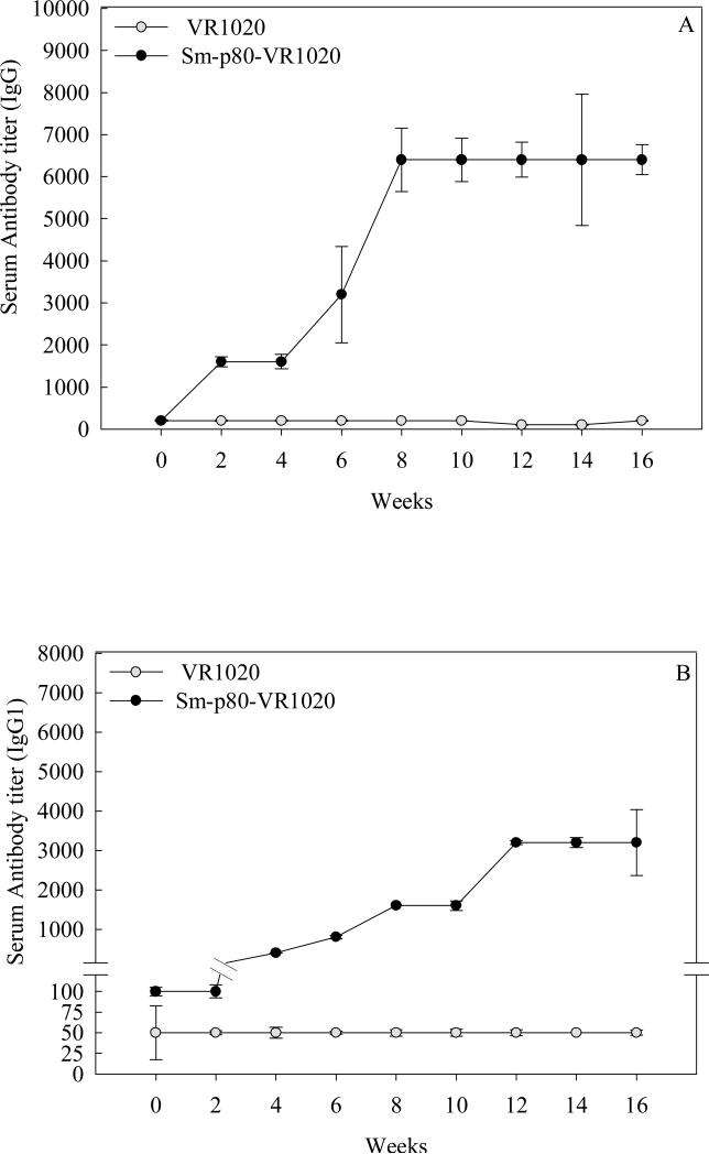
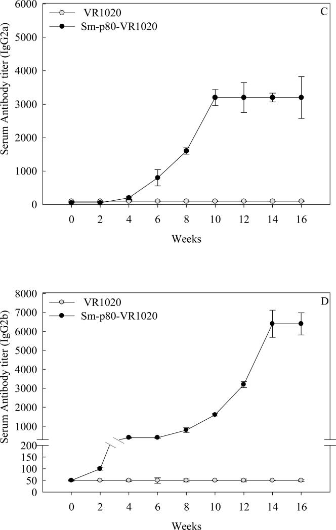
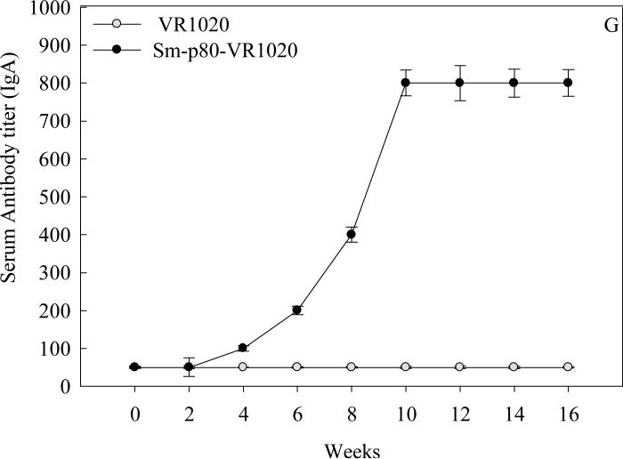
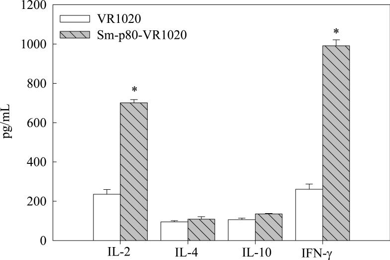
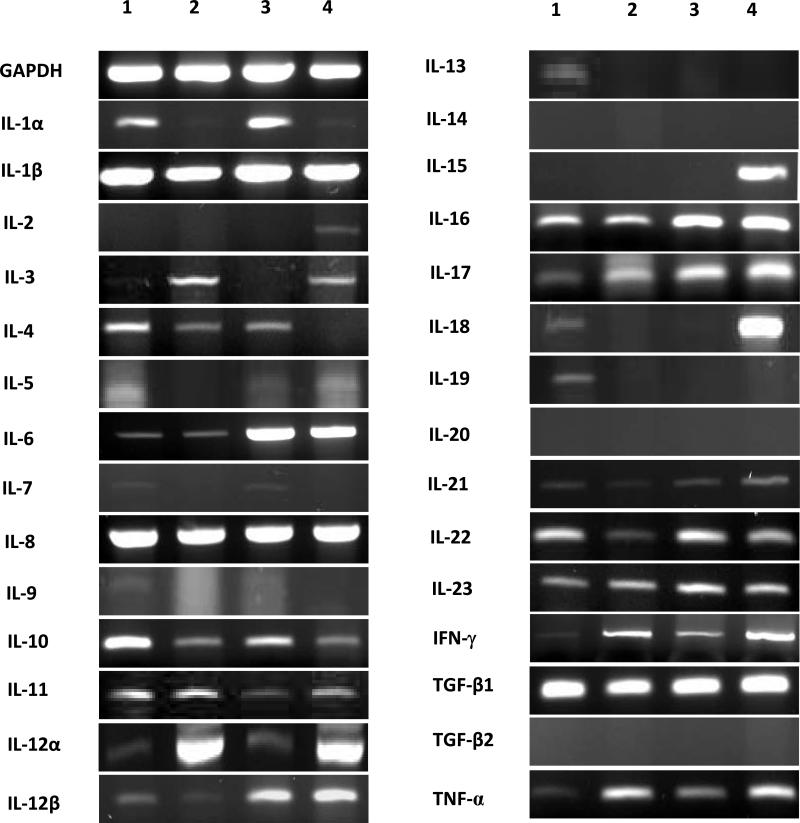
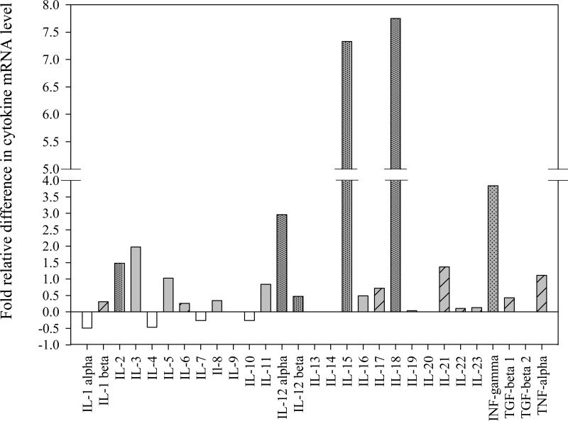
No effective vaccine exists for the human parasitic disease, schistosomiasis. We have targeted a functionally important antigen, Sm-p80 as a vaccine candidate because of its consistent immunogenicity, protective potential and important role in the immune evasion process. In this study we report that a Sm-p80-based DNA vaccine formulation confers 59% reduction in worm burden in mice. Animals immunized with Sm-p80-pcDNA3 exhibited a decrease in egg production by 84%. Sm-p80 DNA elicited strong immune responses that include IgG2A and IgG2B antibody isotypes in vaccinated animals. Splenocytes proliferated in response to Sm-p80 produced appreciably more Th1 response enhancing cytokines (IL-2, IFN-gamma) than Th2 response enhancing cytokines (IL-4, IL-10). These data reinforce the potential of Sm-p80 as an excellent vaccine candidate for schistosomiasis.


Figures

Figure 1
Figure 1
Titers of anti-Sm-p80 antibodies in immunized mice. ELISA was performed with a pool of sera obtained by mixing equal volumes of serum collected from each mouse(biweekly) in their respective groups (VR1020 and Sm-p80-VR1020). IgG (A), IgG1 (B) IgG2a (C), IgG2b (D) IgG3 (E) IgM(F) and IgA (G).The value represent the mean of three experiments ± standard error. Statistic significance (P<0.05) are indicated by (*) compared with VR1020 control group.
Figure 1
Figure 1
Titers of anti-Sm-p80 antibodies in immunized mice. ELISA was performed with a pool of sera obtained by mixing equal volumes of serum collected from each mouse(biweekly) in their respective groups (VR1020 and Sm-p80-VR1020). IgG (A), IgG1 (B) IgG2a (C), IgG2b (D) IgG3 (E) IgM(F) and IgA (G).The value represent the mean of three experiments ± standard error. Statistic significance (P<0.05) are indicated by (*) compared with VR1020 control group.
Figure 2
Figure 2
Levels of cytokine production by splenocytes after 48 hr stimulation with recombinant Sm-p80 in vitro. Groups of mice were inoculated with VR1020 and Sm-p80-VR1020. Data are shown as mean ± S.D. Statistical significance (P<0.05) are indicated by (*) compared with VR1020 control group using independent sample t- test.
Figure 3
Figure 3
RT-PCR for cytokine expression by splenocytes after 24 hr stimulation with recombinant Sm-p80 in vitro. Panels 1, and 3, control groups inoculated with VR1020; Panels 2, 4, experimental groups inoculated with Sm-p80-VR1020. 1, 2, medium control; 3,4, splenocytes stimulated with recombinant Sm-p80.
Figure 4
Figure 4
Fold relative difference of cytokine mRNA level by splenocytes after 24 hr stimulation with recombinant Sm-p80 in vitro. The fold difference was calculated by comparing the differences in the message levels of the control group (VR1020) with the experimental group (Sm-p80-VR1020) after standardization using respective GAPDH.

Similar articles

Cited by

References

    1. Berriman M, Haas BJ, LoVerde PT, et al. The genome of the blood fluke Schistosoma mansoni. Nature. 2009;460:352–358. - PMC - PubMed
    1. Hotez PJ, Molyneux DH, Fenwick A, et al. Control of neglected tropical diseases. N Engl J Med. 2007;357:1018–1027. - PubMed
    1. Steinmann P, Keiser J, Bos R, Tanner M, Utzinger J. Schistosomiasis and water resources development: systematic review, meta-analysis, and estimates of people at risk. Lancet Infect Dis. 2006;6:411–425. - PubMed
    1. Young BW, Podesta RB. Complement and 5-HT increase phosphatidylcholine incorporation into the outer bilayers of Schistosoma mansoni. J Parasitol. 1986;72:802–803. - PubMed
    1. Ahmad G, Torben W, Zang W, Matt W, Siddiqui AA. Sm-p80 based DNA vaccine formulation induces potent protective immunity against Schistosoma mansoni,. Parasite Immunol. 2008;31:156–161. - PMC - PubMed

Publication types