Review
. 2009 Apr;22(2):175-86.
doi: 10.1111/j.1755-148X.2009.00554.x. Epub 2009 Feb 14.

The good and the bad of chemokines/chemokine receptors in melanoma

Affiliations
Review

The good and the bad of chemokines/chemokine receptors in melanoma

Ann Richmond et al. Pigment Cell Melanoma Res. 2009 Apr.

Abstract

Chemokine ligand/receptor interactions affect melanoma cell growth, stimulate or inhibit angiogenesis, recruit leukocytes, promote metastasis, and alter the gene expression profile of the melanoma associated fibroblasts. Chemokine/chemokine receptor interactions can protect against tumor development/growth or can stimulate melanoma tumor progression, tumor growth and metastasis. Metastatic melanoma cells express chemokine receptors that play a major role in the specifying the organ site for metastasis, based upon receptor detection of the chemokine gradient elaborated by a specific organ/tissue. A therapeutic approach that utilizes the protective benefit of chemokines involves delivery of angiostatic chemokines or chemokines that stimulate the infiltration of cytotoxic T cells and natural killer T cells into the tumor microenvironment. An alternative approach that tackles the tumorigenic property of chemokines uses chemokine antibodies or chemokine receptor antagonists to target the growth and metastatic properties of these interactions. Based upon our current understanding of the role of chemokine-mediated inflammation in cancer, it is important that we learn to appropriately regulate the chemokine contribution to the tumorigenic 'cytokine/chemokine storm', and to metastasis.


Figures

Figure 1
Figure 1
Diagramic representation of the family of human chemokines and their receptors. The chemokines are represented by the ligand number and the receptors to which they bind are indicated according to their classification of CXC, C, CX3C or CC. Accordingly the ligand number beside the receptor indicates CC or CXC chemokine. For example, the `8' adjacent to `CXCR1' represents CXCL8. The colors are an indication of different chromosomal localization of the genes for that ligand such that if the color for all the ligands for one receptor is the same, then they are all found on the same chromosome. Two chemokines, CXCL16 and CX3CL1, are transmembrane proteins and the extra line on the ligand circle indicates this. [This figure is reprinted with permission from Zlotnik et al. (2006), Biomed Central.]
Figure 2
Figure 2
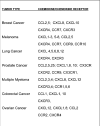
Cancer cells display a variety of chemokine receptors and produce multiple chemokines. This figure lists the chemokine receptors and ligands reported to be produced in a number of types of cancer.
Figure 3
Figure 3
Melanoma tumor cells elaborate CXC and CC chemokines that affect angiogenesis, inflammation, the tumor microenvironment, immune cell response, recruitment of leukocytes, tumor growth and metastasis. The melanoma cells also express chemokine receptors that direct metastasis to specific target organs that elaborate a chemokine gradient that binds and activates these melanoma cell chemokine receptors. Chemokines also stimulate production of metalloproteinases that degrade the matrix and facilitate melanoma metastasis. CXC, CXC chemokine ligands; CXCR, CXC chemokine ligand receptor; CC, CC chemokine ligands; CCR, CC chemokine ligand receptor; EPC, endothelial progenitor cell; MISC, myeloid immune suppressor cells; MMPs, matrix metalloproteinases; VEGF, vascular endothelial growth factor.

Similar articles

Cited by

References

    1. Airoldi I, Di Carlo E, Cocco C, Taverniti G, D'Antuono T, Ognio E, Watanabe M, Ribatti D, Pistoia V. Endogenous IL-12 triggers an antiangiogenic program in melanoma cells. Proc. Natl Acad. Sci. USA. 2007;104:3996–4001. - PMC - PubMed
    1. Amersi FF, Terando AM, Goto Y, Scolyer RA, Thompson JF, Tran AN, Faries MB, Morton DL, Hoon DS. Activation of CCR9/CCL25 in cutaneous melanoma mediates preferential metastasis to the small intestine. Clin. Cancer Res. 2008;14:638–645. - PMC - PubMed
    1. Balentien E, Mufson BE, Shattuck RL, Derynck R, Richmond A. Effects of MGSA/GRO alpha on melanocyte transformation. Oncogene. 1991;6:1115–1124. - PubMed
    1. Balkwill F, Charles KA, Mantovani A. Smoldering and polarized inflammation in the initiation and promotion of malignant disease. Cancer Cell. 2005;7:211–217. - PubMed
    1. Balkwill F, Mantovani A. Inflammation and cancer: back to Virchow? Lancet. 2001;357:539–545. - PubMed

Publication types