. 2009;11(1):R23.
doi: 10.1186/ar2616. Epub 2009 Feb 17.

The proliferative human monocyte subpopulation contains osteoclast precursors

Affiliations

The proliferative human monocyte subpopulation contains osteoclast precursors

Roya Lari et al. Arthritis Res Ther. 2009.

Abstract

Introduction: Immediate precursors of bone-resorbing osteoclasts are cells of the monocyte/macrophage lineage. Particularly during clinical conditions showing bone loss, it would appear that osteoclast precursors are mobilized from bone marrow into the circulation prior to entering tissues undergoing such loss. The observed heterogeneity of peripheral blood monocytes has led to the notion that different monocyte subpopulations may have special or restricted functions, including as osteoclast precursors.

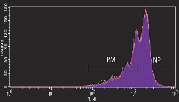
Methods: Human peripheral blood monocytes were sorted based upon their degree of proliferation and cultured in macrophage colony-stimulating factor (M-CSF or CSF-1) and receptor activator of nuclear factor-kappa-B ligand (RANKL).

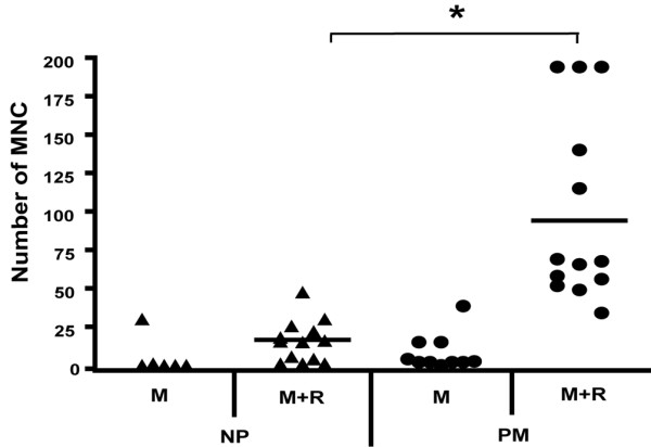
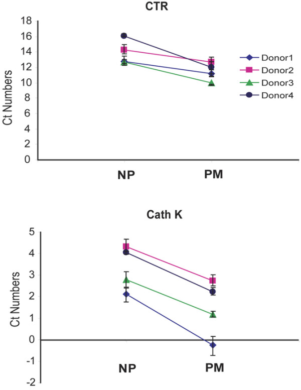
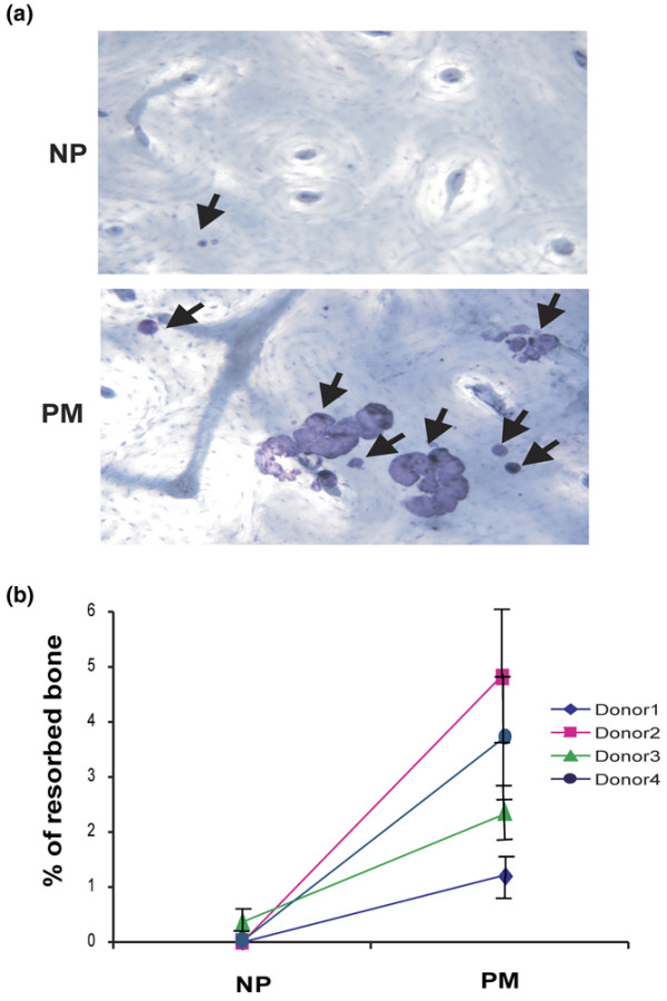
Results: The monocyte subpopulation that is capable of proliferation gave rise to significantly more multinucleated, bone-resorbing osteoclasts than the bulk of the monocytes.

Conclusions: Human peripheral blood osteoclast precursors reside in the proliferative monocyte subpopulation.


Figures

Figure 1
Figure 1
Sorting proliferative monocyte (PM) and non-proliferative (NP) population cells after carboxyfluorescein diacetate-succinimidyl ester (CFSE) labeling and culture. CFSE-labeled peripheral blood mononuclear cells were cultured in alpha-minimum essential medium + 3% heat-inactivated fetal bovine serum containing macrophage colony-stimulating factor (8,000 U/mL) in non-treated dishes for 9 days. The adherent cells were then sorted based on their CFSE fluorescence intensity as PM (CFSElo) and NP (CFSEhi) populations [25].
Figure 2
Figure 2
Proliferative monocytes (PMs) contain precursors of tartrate-resistant acid phosphatase-positive (TRAP+) multinucleated cells (MNCs). Non-proliferative (NP) and PM subpopulations from 13 donors, sorted as in Figure 1, were cultured in duplicate or triplicate cultures in macrophage colony-stimulating factor (M-CSF) (8,000 U/mL) and receptor activator of nuclear factor-kappa-B ligand (RANKL) (50 ng/mL) for 21 days; because insufficient cells were available, the two starting populations from fewer donors were also cultured in M-CSF alone. TRAP+ MNCs were counted. The mean number of such cells was significantly higher in the PM-derived cells cultured in M-CSF and RANKL compared with the NP-derived population (*P < 0.001). M, macrophage colony-stimulating factor; R, receptor activator of nuclear factor-kappa-B ligand.
Figure 3
Figure 3
Osteoclast gene expression in differentiated proliferative monocyte (PM) and non-proliferative (NP) subpopulations. NP and PM subpopulations, sorted as in Figure 1, were cultured for 14 days in macrophage colony-stimulating factor (8,000 U/mL) and receptor activator of nuclear factor-kappa-B ligand (50 ng/mL). Calcitonin receptor (CTR) and cathepsin K (Cath K) mRNA expression were measured by quantitative polymerase chain reaction. Samples from four individual donors were tested in triplicate, and data were normalized to 18S expression for each gene. Values are means of cycle threshold (Ct) numbers that were obtained in each sample ± standard error. The mean values for the PM population were significantly lower than those for the correspondingly treated NP population from the same donor (P ≤ 0.05).
Figure 4
Figure 4
Precursors of bone-resorbing cells reside in the proliferative monocyte (PM) population. Sorted non-proliferative (NP) and PM populations (Figure 1) were cultured on bovine bone (3 × 10⁴ cells per slice) for 21 days in the presence of macrophage colony-stimulating factor (8,000 U/mL) and receptor activator of nuclear factor-kappa-B ligand (50 ng/mL). (a) The bone slices were stained with haematoxylin (magnification × 200). Arrows indicate pits on the bone surface. (b) Resorption pit area measured for four donors (see 'Higher bone resorption in the proliferative monocyte population following culture in M-CSF and RANKL' section). Values are means of percentage of resorbed bone ± standard error. For each donor, the mean values for the PM group are significantly greater than those for the correspondingly treated NP cells (P < 0.05).

Comment in

References

    1. Romas E, Gillespie MT, Martin TJ. Involvement of receptor activator of NF[kappa]B ligand and tumor necrosis factor-[alpha] in bone destruction in rheumatoid arthritis. Bone. 2002;30:340–346. doi: 10.1016/S8756-3282(01)00682-2. - DOI - PubMed
    1. Kong YY, Feige U, Sarosi I, Bolon B, Tafuri A, Morony S, Capparelli C, Li J, Elliott R, McCabe S, Wong T, Campagnuolo G, Moran E, Bogoch ER, Van G, Nguyen LT, Ohashi PS, Lacey DL, Fish E, Boyle WJ, Penninger JM. Activated T cells regulate bone loss and joint destruction in adjuvant arthritis through osteoprotegerin ligand. Nature. 1999;402:304–309. doi: 10.1038/46303. - DOI - PubMed
    1. Fuller K, Wong B, Fox S, Choi Y, Chambers TJ. TRANCE is necessary and sufficient for osteoblast-mediated activation of bone resorption in osteoclasts. J Exp Med. 1998;188:997–1001. doi: 10.1084/jem.188.5.997. - DOI - PMC - PubMed
    1. Lacey DL, Timms E, Tan HL, Kelley MJ, Dunstan CR, Burgess T, Elliott R, Colombero A, Elliott G, Scully S, Hsu H, Sullivan J, Hawkins N, Davy E, Capparelli C, Eli A, Qian YX, Kaufman S, Sarosi I, Shalhoub V, Senaldi G, Guo J, Delaney J, Boyle WJ. Osteoprotegerin ligand is a cytokine that regulates osteoclast differentiation and activation. Cell. 1998;93:165–176. doi: 10.1016/S0092-8674(00)81569-X. - DOI - PubMed
    1. Tanaka S, Takahashi N, Udagawa N, Tamura T, Akatsu T, Stanley ER, Kurokawa T, Suda T. Macrophage colony-stimulating factor is indispensable for both proliferation and differentiation of osteoclast progenitors. J Clin Invest. 1993;91:257–263. doi: 10.1172/JCI116179. - DOI - PMC - PubMed

Publication types

MeSH terms