. 2009 May;29(9):2443-55.
doi: 10.1128/MCB.01443-08. Epub 2009 Feb 17.

Transforming growth factor beta depletion is the primary determinant of Smad signaling kinetics

Affiliations

Transforming growth factor beta depletion is the primary determinant of Smad signaling kinetics

David C Clarke et al. Mol Cell Biol. 2009 May.

Erratum in

Abstract

A cell's decision to growth arrest, apoptose, or differentiate in response to transforming growth factor beta (TGF-beta) superfamily ligands depends on the ligand concentration. How cells sense the concentration of extracellular bioavailable TGF-beta remains poorly understood. We therefore undertook a systematic quantitative analysis of how TGF-beta ligand concentration is transduced into downstream phospho-Smad2 kinetics, and we found that the rate of TGF-beta ligand depletion is the principal determinant of Smad signal duration. TGF-beta depletion is caused by two mechanisms: (i) cellular uptake of TGF-beta by a TGF-beta type II receptor-dependent mechanism and (ii) reversible binding of TGF-beta to the cell surface. Our results indicate that cells sense TGF-beta dose by depleting TGF-beta via constitutive TGF-beta type II receptor trafficking processes. Our results also have implications for the role of the TGF-beta type II receptor in disease, as tumor cells harboring TGF-beta type II receptor mutations exhibit impaired TGF-beta depletion, which may contribute to the overproduction of TGF-beta and a consequently poor prognosis in cancer.


Figures
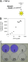
FIG. 1.
FIG. 1.
Ligand concentration specifies the cellular responses to TGF-β. (A) Schematic of the luciferase reporter assay. Cells possess a stably integrated luciferase reporter gene driven by a Smad-sensitive promoter. Addition of TGF-β induces production of luciferase, which catalyzes a light-producing reaction that can be measured with a luminometer. (B) Luciferase reporter gene activity in PE25 mink lung epithelial cells varies in a sigmoidal manner to the log10 concentration of TGF-β. (C) The growth inhibitory response to TGF-β in PE25 cells is concentration dependent. PE25 cells were plated at a low density and grown in the presence of 0, 1, 5, 10, 25, or 50 pM TGF-β for 2 weeks followed by crystal violet staining. Cells were unable to proliferate in the presence of 5 pM or more of TGF-β.
FIG. 2.
FIG. 2.
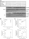
The number of TGF-β molecules per cell predicts the Smad signal. (A) Immunoblots of the indicated proteins in whole-cell lysates of PE25 cells exposed under the indicated conditions. (B and C) Plots of the ratio between the immunoblot band densities of phospho-Smad2 (P-Smad2) and α-tubulin versus TGF-β concentration (pM) (B) or the calculated number of TGF-β molecules per cell (C) at 30 min and 8 h. Note the reduced spread of the data points in panel C compared to those in panel B.
FIG. 3.
FIG. 3.
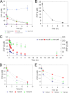
TGF-β is depleted from the culture medium during TGF-β signaling. (A) The TGF-β reporter assay specifically measures TGF-β in conditioned medium. PE25 cells were exposed to either fresh medium or 25 pM TGF-β for 0.5, 4, and 8 h, after which the culture medium was transferred to the reporter cells. The following treatments were applied at the time of transfer: untreated control (25 pM), addition of 25 pM TGF-β to the cultured medium (spike TGF-β), and addition of SB-431542 or vehicle (dimethyl sulfoxide [DMSO]). The fresh medium group showed no increase in luciferase activity above background over time, indicating that the PE25 cells do not secrete bioactive TGF-β under normal conditions. Spiking additional TGF-β into the transferred medium induced reporter activity to saturated levels, demonstrating that the reporter cells remain competent to respond to TGF-β during the assay. Adding SB-431542 caused a decrease in the luciferase activity to basal levels, whereas the DMSO vehicle did not, indicating that TGF-β is specifically causing the increase in reporter cell luciferase activity. We conclude that our reporter assay specifically measures bioactive TGF-β in the culture medium. (B) External validation of the TGF-β reporter assay. An ELISA specific to human TGF-β1 was used to measure TGF-β levels in the culture medium of PE25 cells exposed to an initial dose of 25 pM TGF-β for 0.5, 4, and 8 h. The ELISA revealed the same depletion kinetics as observed using the TGF-β reporter assay, thus externally validating the reporter assay. (C) TGF-β depletion kinetics. PE25 cells were exposed to 10, 25, or 200 pM TGF-β (corresponding to 6,020, 15,050, and 120,400 TGF-β molecules per cell, respectively, for the experimental conditions) and TGF-β concentration in the culture medium was measured at the indicated times by the TGF-β reporter assay. Data points are the averages of three independent experiments and error bars represent the standard deviations. (D) TGF-β is depleted by several TGF-β-responsive cell types. A 25 pM dose of TGF-β was applied to the indicated cell types for 8 h and TGF-β concentration in the culture medium was measured using the TGF-β reporter assay at 0.5, 4, and 8 h after the initial dose was applied. (E) Three human TGF-β isoforms are depleted by PE25 cells. A 25 pM concentration of both TGF-β2 and TGF-β3 was applied to PE25 cells for 8 h and the TGF-β concentration in the culture medium was measured using the TGF-β reporter assay at 0.5, 4, and 8 h after the initial dose was applied. The data for the TGF-β1 isoform were taken from panel C for comparison. In both panels B and C, the data points are the average TGF-β concentrations and the error bars represent the standard deviations of three replicate measurements.
FIG. 4.
FIG. 4.
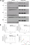
Phospho-Smad2 time course as a function of TGF-β dose. (A) Raw immunoblot assay data, showing phospho-Smad2 (P-Smad2) levels at the indicated times in response to 10, 25, and 200 pM TGF-β. In each gel, known amounts of the phosphorylated recombinant MH2 domain (P-MH2) spiked into cell lysates treated with 100 pM TGF-β were run alongside the time course samples in order to estimate the number of phospho-Smad2 molecules per cell in these samples. The order in which the standards were loaded on the gel was arranged to minimize overlap of the signals such that the density of each band could be accurately quantified. Smad2 immunoblots for the time courses are also shown for each dose, showing that Smad2 is constitutively present during signaling. (B) Interpolated values of phospho-Smad2 molecules per cell are plotted versus time. Data points represent the averages and error bars represent the standard deviations of three independent experiments. Note that the large error bars reflect mainly quantitative differences between gels, as the qualitative trends in the data were reproducible. In other words, the curves for each dose were qualitatively similar but were shifted in relation to each other along the ordinate, thus conferring sizeable error bars. (C) Time course of Smad nuclear activity in response to different doses of TGF-β. PEZS cells were exposed to either 10, 25, or 200 PM TGF-β for the indicated times, at which point luciferase reporter activity was measured. Luciferase activities from TGF-β-treated cells were normalized to values from untreated control cells.
FIG. 5.
FIG. 5.
TGF-β depletion is mediated by a TβRII-dependent mechanism and reversible binding to the cell surface. (A) The TβRII, but not the TβRI, is necessary for TGF-β depletion. TGF-β depletion time courses (25 pM initial dose) were measured for PE25 (representative of the Mv1Lu parental cell line), R1B (lack functional TβRI), and DR27 (lack functional TβRII) cells. Depletion was impaired in DR27 but not R1B cells compared to PE25 cells. (B) Detailed kinetics of TGF-β depletion by DR27 cells. The experiment was carried out in an identical manner to that shown in panel A except that the TGF-β concentration was measured at additional time points for the DR27 cells. (C) TGF-β depletion kinetics in DR27 cells are consistent with a mechanism involving reversible binding to the cell surface. A washout experiment was performed in which DR27 and PE25 cells were exposed to TGF-β for 60 min, after which the medium was replaced with fresh medium containing no TGF-β. TGF-β concentration was then measured by reporter assay at the indicated times. (D) TGF-β depletion is impaired at 4°C. TGF-β depletion time courses (25 pM initial dose) were measured for PE25 cells at either 4°C or 37°C. In panels A, B, and D, data points are the averages and error bars are the standard deviations for three replicates.
FIG. 6.
FIG. 6.
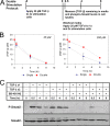
TGF-β receptor levels are preserved during signaling. (A) Design of the double-stimulation experiment. Two groups of cells were seeded for comparison: a single-stimulation group and a double-stimulation group. First, the indicated dose of TGF-β was applied to the double-stimulation cells for 8 h while fresh medium lacking TGF-β was applied the single-stimulation group. At 8 h, the media of both groups were washed out, followed by application of 25 pM TGF-β for another 8 h, during which the TGF-β depletion from the culture medium and corresponding phospho-Smad2 levels were measured at the indicated times for both groups. (B) TGF-β concentrations remaining in the culture media of cells during the second 8 h of the double-stimulation experiment. The depletion rates of an initial dose of 25 pM TGF-β were similar for the single- and double-stimulation groups, regardless of whether the double-stimulation group was preexposed to 25 or 200 pM TGF-β. (The increase of ∼10 pM TGF-β in the 200 pM double-stimulation group reflects dissociation of reversibly bound TGF-β that did not get removed during the washout.) (C) Phospho-Smad2 immunoblots of the lysates of cells in the double-stimulation experiment. A second dose of 25 pM TGF-β restored phospho-Smad2 levels in cells preexposed to 25 pM TGF-β. Applying the small-molecule inhibitor to the TβRI (SB-431542) for the second 8 h eliminated the phospho-Smad2 signals in both the single- and double-stimulation groups. (The faint band observed at 0.5 h for the TGF-βx2/SB-431542 group is phospho-Smad2 remaining from the first dose of TGF-β that was applied prior to the addition of SB-431542.) In addition, Smad2 levels were preserved throughout the duration of the experiment.
FIG. 7.
FIG. 7.
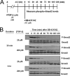
Phospho-Smad2 is dephosphorylated at a similar rate throughout signaling. (A) PE25 cells were exposed to either 25 or 200 pM TGF-β for either 30 min or 6 h and then exposed to 10 μM TβRI inhibitor SB-431542. Phospho-Smad2 (P-Smad2) and Smad2 in whole-cell lysates was measured by immunoblotting every 15 min for 2 h after applying the TβRI inhibitor. (B) Phospho-Smad2 decreased to basal levels by about 60 min in each case, whereas Smad2 levels did not change appreciably. Cntl, untreated control.
FIG. 8.
FIG. 8.
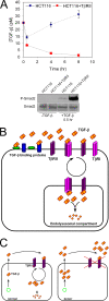
TβRII mutations in cancer impair TGF-β depletion. (A) TGF-β depletion is impaired in HCT116 cells and restored upon expression of the TβRII. Cells were exposed to a 25 pM concentration of TGF-β for 8 h, with TGF-β levels remaining in the culture medium measured at 0.5, 4, and 8 h after the dose was applied. Data points are the averages of three replicates and the error bars represent the standard deviations. Phospho-Smad2 and Smad2 levels in the cell lysates from the 0- and 0.5-h time points from the above experiment were measured by immunoblotting (lower panel). (B) Model of TGF-β depletion processes. (C) Model of how normal and cancer cells regulate the extracellular levels of TGF-β. TGF-β levels in the extracellular fluids are maintained by the balance between cellular production/secretion of TGF-β and depletion mediated by the TβRII. Cancer cells often overproduce TGF-β and those cell types lacking the TβRII would also be unable to deplete TGF-β, thus exacerbating the rise of TGF-β levels in the extracellular fluids, which correlates with poor prognosis.

References

    1. Abe, M., J. G. Harpel, C. N. Metz, I. Nunes, D. J. Loskutoff, and D. B. Rifkin. 1994. An assay for transforming growth factor-beta using cells transfected with a plasminogen activator inhibitor-1 promoter-luciferase construct. Anal. Biochem. 216276-284. - PubMed
    1. Afrakhte, M., A. Moren, S. Jossan, S. Itoh, K. Sampath, B. Westermark, C. H. Heldin, N. E. Heldin, and P. ten Dijke. 1998. Induction of inhibitory Smad6 and Smad7 mRNA by TGF-β family members. Biochem. Biophys. Res. Commun. 249505-511. - PubMed
    1. Ashe, H. L., and J. Briscoe. 2006. The interpretation of morphogen gradients. Development 133385-394. - PubMed
    1. Bangi, E., and K. Wharton. 2006. Dual function of the Drosophila Alk1/Alk2 ortholog Saxophone shapes the Bmp activity gradient in the wing imaginal disc. Development 1333295-3303. - PubMed
    1. Belenkaya, T. Y., C. Han, D. Yan, R. J. Opoka, M. Khodoun, H. Liu, and X. Lin. 2004. Drosophila Dpp morphogen movement is independent of dynamin-mediated endocytosis but regulated by the glypican members of heparan sulfate proteoglycans. Cell 119231-244. - PubMed

Publication types

MeSH terms

Substances

LinkOut - more resources