. 2009 Apr;29(8):2155-67.
doi: 10.1128/MCB.01325-08. Epub 2009 Feb 17.

Identification of a cytoplasmic complex that adds a cap onto 5'-monophosphate RNA

Affiliations

Identification of a cytoplasmic complex that adds a cap onto 5'-monophosphate RNA

Yuichi Otsuka et al. Mol Cell Biol. 2009 Apr.

Abstract

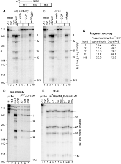
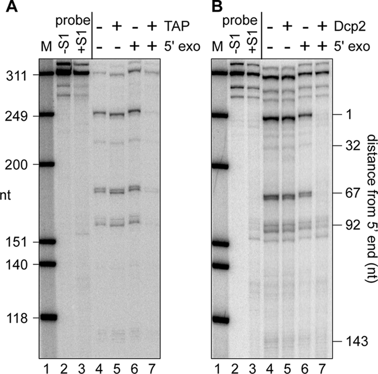
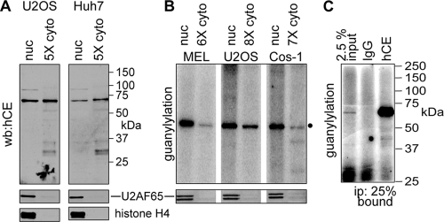
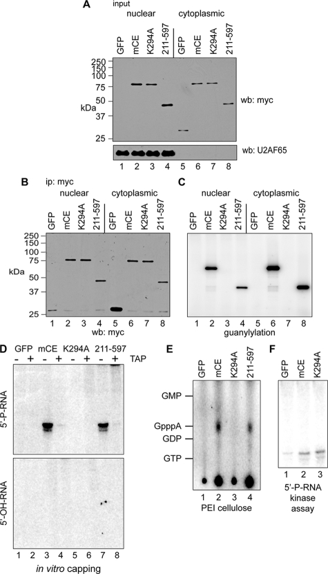
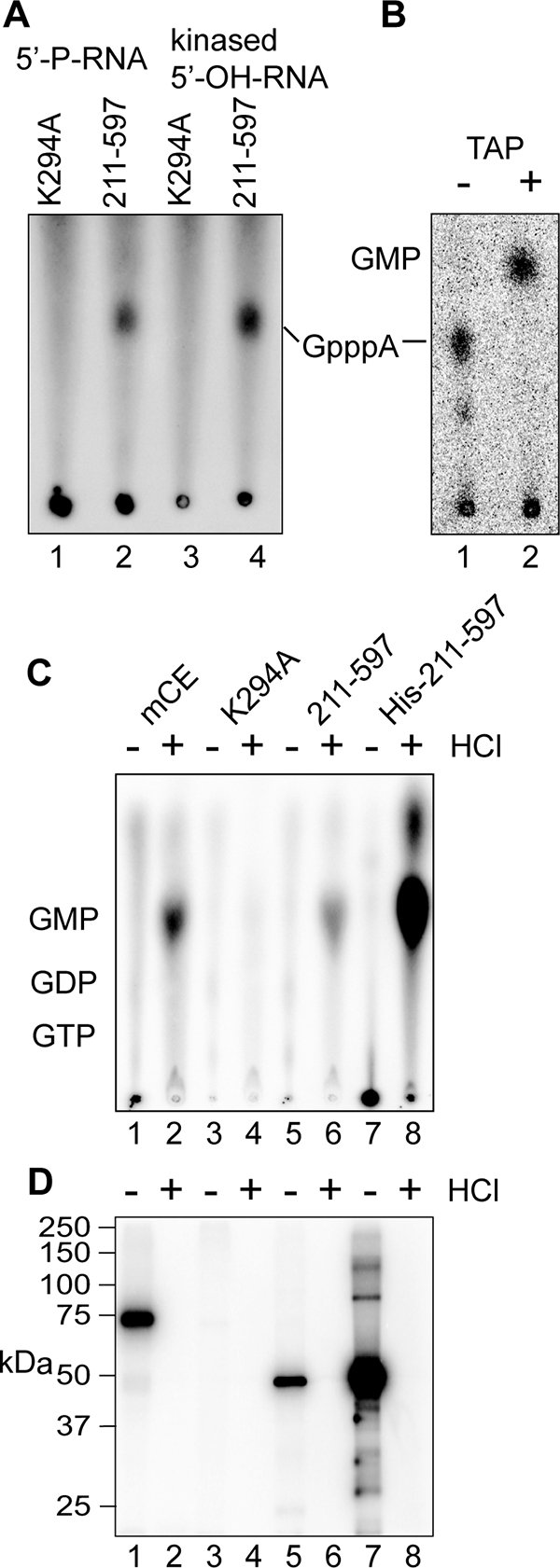
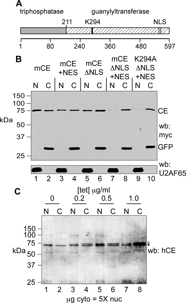
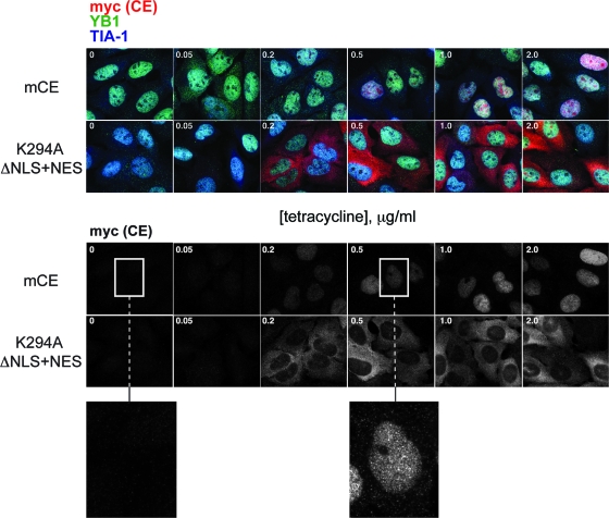
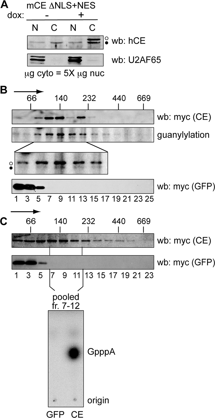
Endonuclease decay of nonsense-containing beta-globin mRNA in erythroid cells generates 5'-truncated products that were reported previously to have a cap or caplike structure. We confirmed that this 5' modification is indistinguishable from the cap on full-length mRNA, and Western blotting, immunoprecipitation, and active-site labeling identified a population of capping enzymes in the cytoplasm of erythroid and nonerythroid cells. Cytoplasmic capping enzyme sediments in a 140-kDa complex that contains a kinase which, together with capping enzyme, converts 5'-monophosphate RNA into 5'-GpppX RNA. Capping enzyme shows diffuse and punctate staining throughout the cytoplasm, and its staining does not overlap with P bodies or stress granules. Expression of inactive capping enzyme in a form that is restricted to the cytoplasm reduced the ability of cells to recover from oxidative stress, thus supporting a role for capping in the cytoplasm and suggesting that some mRNAs may be stored in an uncapped state.


Figures

FIG. 1.
FIG. 1.
Recovery of decay intermediates with trimethyl cap antibody and eIF4E. (A) A total of 40 μg of cytoplasmic RNA from Thal10 cells was incubated with H20 cap antibody bound to protein G-Sepharose. The recovered beads were washed twice with binding buffer (lanes 5 and 6), twice with 50 μM GDP (lanes 7 and 8), and eluted twice with 50 μM m7GDP (lanes 9 and 10). RNA in each eluate was recovered by ethanol precipitation and analyzed by S1 nuclease protection assay using a 5′-labeled antisense probe (top) from nucleotide 250 past the 5′ end of β-globin mRNA. As a control, 8 μg of Thal10 cell cytoplasmic RNA was analyzed in lane 3 (input). A sample of the S1 probe plus 10 μg of yeast tRNA is in lane 1 (probe, −S1), and the same sample after S1 nuclease digestion is in lane 2 (probe, +S1). The positions of HinfI restriction fragments of ΦX174 DNA electrophoresed on the gel (molecular size markers) are indicated on the left, and the identified β-globin mRNA decay intermediates are indicated on the right. (B) Cytoplasmic RNA from Thal10 cells was analyzed as in panel A, with the exception that the RNA was recovered with GST-eIF4E bound to glutathione-Sepharose. (C) The relative amounts of full-length mRNA and each decay intermediate recovered in panels A and B were quantified by a PhosphorImager, and the results were normalized to full-length mRNA and each decay intermediate in the input RNA. (D) Cytoplasmic RNA from Thal10 cells was bound onto immobilized H20 cap antibody, washed as in panel A, and eluted with 50 μM GDP (lane 4) or the indicated concentrations of m7GDP (lanes 5, 6, and 7). (E) Cytoplasmic RNA from Thal10 cells was incubated with immobilized eIF4E in the presence of 0, 0.1, 1, 10, or 100 μM m7GpppG (lanes 5 to 8) or ApppG (lanes 9 to 12). Bound complexes were washed with binding buffer containing 50 μM GDP, eluted with 50 μM m7GDP, and analyzed by S1 nuclease protection assay. The positions of full-length mRNA and each of the decay intermediates are indicated on the right. As a control, 20% of Thal10 cell cytoplasmic RNA was applied to lane 3 (input).
FIG. 2.
FIG. 2.
Susceptibility of full-length β-globin mRNA and decay intermediates to cap hydrolysis and cap analog competition for binding to eIF4E and cap antibody. Cytoplasmic RNA from Thal10 cells was treated without (−) (lanes 4 and 6) or with (+) (lanes 5 and 7) TAP (A) or Dcp2 (B). RNA recovered from each reaction was then incubated without (lanes 4 and 5) or with (lanes 6 and 7) a 5′-monophosphate-dependent 5′ exonuclease and analyzed by S1 nuclease protection assay.
FIG. 3.
FIG. 3.
Biochemical evidence for cytoplasmic capping enzyme. (A) The indicated relative amounts of nuclear and cytoplasmic extracts of U2OS and Huh7 cells were analyzed by Western blotting (wb) with an affinity-purified sheep antibody against human capping enzyme (hCE). To control leakage of capping enzyme during cell lysis, the same samples were analyzed by Western blotting with antibodies to U2AF65 and histone H4 (lower panels). (B) The guanylylation activity of capping enzyme present in the indicated amounts of nuclear and cytoplasmic extracts from MEL cells, U2OS cells, and COS-1 cells was determined by incubating with [α-32P]GTP, followed by electrophoresis on a 10% SDS-PAGE gel and PhosphorImager analysis. In the lower panel, the same extracts were analyzed by Western blotting with antibody to U2AF65. (C) Cytoplasmic extract from COS-1 cells in panel B was immunoprecipitated with nonimmune IgG or antibody to human capping enzyme. A total of 25% of the immunoprecipitates and 2.5% of input protein were analyzed by guanylylation assay.
FIG. 4.
FIG. 4.
In vitro capping of 5′-monophosphate RNA by immunoprecipitated cytoplasmic capping enzyme. (A) COS-1 cells were transfected with pcDNA3 expressing myc-tagged GFP (lanes 1 and 5), full-length mCE (lanes 2 and 6), mCE with the active-site K294A mutation (lanes 3 and 7), or the guanylylation domain (211-597) alone (lanes 4 and 8). Nuclear and cytoplasmic extracts were assayed by Western blotting with antibodies to the myc tag (top) or to U2AF65 (bottom). (B) Nuclear and cytoplasmic extracts from panel A were immunoprecipitated with immobilized anti-myc monoclonal antibody, and a portion was analyzed by Western blotting with an antibody to the myc tag. (C) Anti-myc antibody-containing beads with the remaining immunoprecipitated protein from panel B were incubated with [α-32P]GTP. A portion was removed after being washed to remove unincorporated GTP and separated on an SDS-PAGE gel, and the guanylylated proteins were visualized by a PhosphorImager. (D) The remaining beads from panel C were incubated with ATP and a 23-nt RNA with a 5′-monophosphate end (5′-P-RNA; top) or 5′-hydroxyl (5′-OH-RNA; bottom). The recovered RNA was treated with (+) or without (−) TAP and visualized by PhosphorImager analysis after electrophoresis on a 15% polyacrylamide/urea gel. (E) RNAs recovered from the reaction with 5′-P-RNA in panel D were digested with P1 nuclease and separated by PEI-cellulose TLC. The mobility of unlabeled nucleotide standards is indicated on the right side of the autoradiograph. (F) Anti-myc antibody-containing beads containing immunoprecipitated GFP, mCE, or K294A from panel B were incubated with 5′-P-RNA and [γ-32P]ATP. The recovered RNA was then electrophoresed on a 15% polyacrylamide/urea gel and visualized by a PhosphorImager.
FIG. 5.
FIG. 5.
Identification of GMP as the nucleoside that is transferred from capping enzyme onto 5′-monophosphate RNA. (A) The 5′-OH-RNA used in Fig. 4D was phosphorylated by incubation with T4 polynucleotide kinase and ATP (kinased 5′-OH-RNA). This and the HPLC-purified 5′-P-RNA used in Fig. 4D were incubated as in that experiment with immunoprecipitated K294A or the 211-597 guanylylation domain plus [α-32P]GTP and ATP. The products recovered by ethanol precipitation were digested with P1 nuclease and separated by PEI-cellulose TLC, as in Fig. 4E. (B) Kinased 5′-OH-RNA was incubated as in panel A except with immunoprecipitated full-length mCE. The RNA recovered by ethanol precipitation was digested with P1 nuclease and analyzed without further treatment on PEI-cellulose (lane 1, −), or treated with TAP prior to TLC (lane 2, +). (C and D) Full-length mCE (lanes 1 and 2), K294A (lanes 3 and 4), the 211 to 597 guanylylation domain (lanes 5 and 6), or a His-tagged recombinant form of the 211 to 597 guanylylation domain expressed in E. coli was immunoprecipitated from U2OS cells expressing each form of the enzyme (lanes 1 to 6) using beads containing anti-myc monoclonal antibody or bound onto Ni-nitrilotriacetic acid agarose (lanes 7 and 8). The immobilized proteins were incubated with [α-32P]GTP, as in Fig. 4C. After being washed to remove unbound GTP, the odd-numbered samples were adjusted to 0.1 N HCl, the even-numbered samples received water, and both were heated for 15 min at 70°C. (C) The acid-treated samples were then neutralized and the supernatants were applied directly to PEI-cellulose and separated as in panel A. (D) SDS sample buffer was added to the beads, and the guanylylated proteins recovered after being heated were separated by SDS-PAGE and visualized by a PhosphorImager.
FIG. 6.
FIG. 6.
Development of forms of capping enzyme that are restricted to the cytoplasm. (A) The organization of mammalian capping enzyme is shown with the location of the guanylylation site (K294) and the NLS highlighted. (B) U2OS cells were transfected with plasmids expressing wild-type mCE (lanes 1 and 2), mCE with addition of the HIV Rev NES (mCE +NES; lanes 3 and 4), mCE with four amino acids deleted from the NLS (mCE ΔNLS; lanes 5 and 6), mCE ΔNLS+NES (lanes 7 and 8), and K294A ΔNLS+NES (lanes 9 and 10). Each of these proteins has an N-terminal myc tag and was cotransfected with a plasmid expressing myc-tagged GFP. Nuclear and cytoplasmic extracts were analyzed as in Fig. 3 by Western blotting with antibodies to the myc tag (top) and U2AF65 (bottom). (C) A stable line of tetracycline-inducible U2OS cells was generated that expresses mCE ΔNLS+NES with an N-terminal myc tag and a C-terminal FLAG tag. These were cultured overnight with the indicated concentrations of tetracycline, and nuclear (N) and cytoplasmic (C) extracts were analyzed by Western blotting (wb) with an affinity-purified rabbit anti-human capping enzyme antibody (hCE). Five times more cytoplasmic extract (on a μg of protein basis) than nuclear extract was applied to the gel. Endogenous capping enzyme is identified with a filled circle, and the slightly larger epitope-tagged protein is identified with an open circle.
FIG. 7.
FIG. 7.
Tetracycline-inducible lines of U2OS cells expressing wild-type mCE or the K294A ΔNLS+NES were cultured for 8 h in medium containing 0, 0.05, 0.2, 0.5, 1, and 2 μg/ml of tetracycline. Fixed cells were stained with antibodies to the myc tag on each form of capping enzyme (CE) (red), YB1 (green), and TIA-1 (blue). The merged micrographs are shown in the upper two panels, and the lower two panels show only the distribution of capping enzyme. Enlargements (3×) of the indicated areas of cells cultured without tetracycline or with 0.5 μg/ml tetracycline are shown beneath.
FIG. 8.
FIG. 8.
Identification of a cytoplasmic capping enzyme complex. (A) U2OS cells stably expressing mCE ΔNLS+NES were cultured for 24 h in the absence (−) or presence (+) of doxycycline (dox), and nuclear (N) and a fivefold-greater amount of cytoplasmic (C) protein was analyzed by Western blotting (wb) with affinity-purified capping enzyme antibody (top) or antibody to U2AF65 (bottom). The open circles correspond to recombinant protein and the filled circles correspond to endogenous capping enzyme. (B) Doxycycline was added to induce recombinant protein expression in cell lines stably transfected with tetracycline-inducible forms of mCE ΔNLS+NES or GFP. Cells were treated with 0.25% formaldehyde for 15 min prior to lysis, and cytoplasmic extracts were separated on linear 10 to 40% glycerol gradients. The sedimentation of mCE ΔNLS+NES or GFP was determined by Western blotting of individual fractions with antibody to the myc tag on each of these proteins, and the cosedimentation of mCE ΔNLS+NES (middle, open circle) with endogenous capping enzyme (CE) (filled circle) was determined by guanylylation assay. The lower panel is an enlargement of that portion of the gradient containing the 140-kDa capping enzyme complex. The positions of MW standards that were run in parallel gradients are indicated on the top panel, with the arrow indicating the direction of sedimentation. (C) mCE ΔNLS+NES and GFP were induced as in panel B by addition of tetracycline to lines of U2OS cells expressing each of these proteins. Cytoplasmic extracts were separated on 10 to 40% glycerol gradients, and capping enzyme and GFP were identified by Western blotting of individual fractions using antibody to the myc tag on each of these proteins. The fractions corresponding to the 140-kDa complex from panel B were pooled and recovered using beads containing immobilized anti-myc monoclonal antibody. These were incubated with [α-32P]GTP, ATP, and 5′-P-RNA, and RNA recovered from this reaction was digested with P1 nuclease and separated on PEI-cellulose TLC to identify the product of the capping reaction.
FIG. 9.
FIG. 9.
Impact of expressing cytoplasmic forms of capping enzyme on cellular recovery from stress. Cultures of U2OS cells stably transfected with plasmids expressing tetracycline-inducible GFP or K294A ΔNLS+NES (left) or GFP or mCE ΔNLS+NES (right) were cultured overnight without (−) or with (+) 2 μg/ml of doxycycline (Dox). The next day, one-half of each set was exposed for 40 min to 0.5 mM sodium arsenite, then washed, and placed into fresh medium. Cell viability was determined 24 h later using the CellTiter-Glo luminescent cell viability assay. Each bar represents the mean ± standard deviation for triplicate samples.

References

    1. Bhattacharyya, S. N., R. Habermacher, U. Martine, E. I. Closs, and W. Filipowicz. 2006. Relief of microRNA-mediated translational repression in human cells subjected to stress. Cell 1251111-1124. - PubMed
    1. Bremer, K. A., A. Stevens, and D. R. Schoenberg. 2003. An endonuclease activity similar to Xenopus PMR1 catalyzes the degradation of normal and nonsense-containing human beta-globin mRNA in erythroid cells. RNA 91157-1167. - PMC - PubMed
    1. Cho, E. J., T. Takagi, C. R. Moore, and S. Buratowski. 1997. mRNA capping enzyme is recruited to the transcription complex by phosphorylation of the RNA polymerase II carboxy-terminal domain. Genes Dev. 113319-3326. - PMC - PubMed
    1. Cougot, N., S. Babajko, and B. Seraphin. 2004. Cytoplasmic foci are sites of mRNA decay in human cells. J. Cell Biol. 16531-40. - PMC - PubMed
    1. Eberle, A. B., S. Lykke-Andersen, O. Muhlemann, and T. H. Jensen. 2009. SMG6 promotes endonucleolytic cleavage of nonsense mRNA in human cells. Nat. Struct. Mol. Biol. 1649-55. - PubMed

Publication types