

Dynamic contrast-enhanced magnetic resonance imaging as a biomarker for prediction of radiation-induced neurocognitive dysfunction
- PMID: 19223506
- PMCID: PMC2699596
- DOI: 10.1158/1078-0432.CCR-08-1420
Dynamic contrast-enhanced magnetic resonance imaging as a biomarker for prediction of radiation-induced neurocognitive dysfunction
Abstract
Purpose: To determine whether early assessment of cerebral microvessel injury can predict late neurocognitive dysfunction after brain radiation therapy (RT).
Experimental design: Ten patients who underwent partial brain RT participated in a prospective dynamic contrast-enhanced magnetic resonance imaging (MRI) study. Dynamic contrast-enhanced MRI was acquired prior to, at weeks 3 and 6 during, and 1 and 6 months after RT. Neuropsychological tests were done pre-RT and at the post-RT MRI follow-ups. The correlations between early delayed changes in neurocognitive functions and early changes in vascular variables during RT were analyzed.
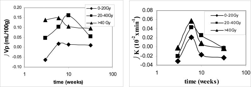
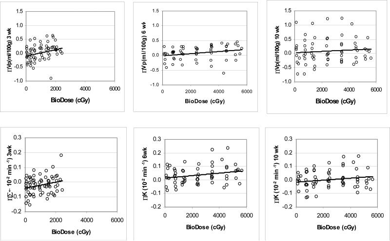
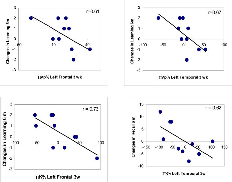
Results: No patients had tumor progression up to 6 months after RT. Vascular volumes and blood-brain barrier (BBB) permeability increased significantly in the high-dose regions during RT by 11% and 52% (P<0.05), respectively, followed by a decrease after RT. Changes in both vascular volume and BBB permeability correlated with the doses accumulated at the time of scans at weeks 3 and 6 during RT and 1 month after RT (P<0.03). Changes in verbal learning scores 6 months after RT were significantly correlated with changes in vascular volumes of left temporal (P<0.02) and frontal lobes (P<0.03), and changes in BBB permeability of left frontal lobes during RT (P<0.007). A similar correlation was found between recall scores and BBB permeability.
Conclusion: Our data suggest that the early changes in cerebral vasculature may predict delayed alterations in verbal learning and total recall, which are important components of neurocognitive function. Additional studies are required for validation of these findings.
Figures
Similar articles
-
A Radiation-Induced Hippocampal Vascular Injury Surrogate Marker Predicts Late Neurocognitive Dysfunction.Int J Radiat Oncol Biol Phys. 2015 Nov 15;93(4):908-15. doi: 10.1016/j.ijrobp.2015.08.014. Epub 2015 Aug 8. Int J Radiat Oncol Biol Phys. 2015. PMID: 26530761 Free PMC article.
-
Use of magnetic resonance imaging to assess blood-brain/blood-glioma barrier opening during conformal radiotherapy.J Clin Oncol. 2005 Jun 20;23(18):4127-36. doi: 10.1200/JCO.2005.07.144. J Clin Oncol. 2005. PMID: 15961760
-
Diffusion tensor imaging of normal-appearing white matter as biomarker for radiation-induced late delayed cognitive decline.Int J Radiat Oncol Biol Phys. 2012 Apr 1;82(5):2033-40. doi: 10.1016/j.ijrobp.2011.01.068. Epub 2011 May 11. Int J Radiat Oncol Biol Phys. 2012. PMID: 21570218 Free PMC article.
-
Neurocognitive Deficits After Radiation Therapy for Brain Malignancies.Am J Clin Oncol. 2015 Dec;38(6):634-40. doi: 10.1097/COC.0000000000000158. Am J Clin Oncol. 2015. PMID: 25503433 Review.
-
Long-term neurocognitive and other side effects of radiotherapy, with or without chemotherapy, for glioma.Cochrane Database Syst Rev. 2019 Aug 5;8(8):CD013047. doi: 10.1002/14651858.CD013047.pub2. Cochrane Database Syst Rev. 2019. PMID: 31425631 Free PMC article.
Cited by
-
Dynamics of rabbit brain edema in focal lesion and perilesion area after traumatic brain injury: a MRI study.J Neurotrauma. 2012 Sep 20;29(14):2413-20. doi: 10.1089/neu.2010.1510. Epub 2011 Oct 4. J Neurotrauma. 2012. PMID: 21675826 Free PMC article.
-
Neurocognitive Dysfunction in Hematopoietic Cell Transplant Recipients: Expert Review from the Late Effects and Quality of Life Working Committee of the Center for International Blood and Marrow Transplant Research and Complications and Quality of Life Working Party of the European Society for Blood and Marrow Transplantation.Biol Blood Marrow Transplant. 2018 Feb;24(2):228-241. doi: 10.1016/j.bbmt.2017.09.004. Epub 2017 Sep 20. Biol Blood Marrow Transplant. 2018. PMID: 28939455 Free PMC article. Review.
-
Blood-brain barrier permeability following conventional photon radiotherapy - A systematic review and meta-analysis of clinical and preclinical studies.Clin Transl Radiat Oncol. 2022 May 4;35:44-55. doi: 10.1016/j.ctro.2022.04.013. eCollection 2022 Jul. Clin Transl Radiat Oncol. 2022. PMID: 35601799 Free PMC article. Review.
-
A pilot study using dynamic contrast enhanced-MRI as a response biomarker of the radioprotective effect of memantine in patients receiving whole brain radiotherapy.Oncotarget. 2016 Aug 9;7(32):50986-50996. doi: 10.18632/oncotarget.9653. Oncotarget. 2016. PMID: 27248467 Free PMC article. Clinical Trial.
-
An Overdetermined System of Transform Equations in Support of Robust DCE-MRI Registration With Outlier Rejection.Tomography. 2016 Sep;2(3):188-196. doi: 10.18383/j.tom.2016.00145. Tomography. 2016. PMID: 28367502 Free PMC article.
References
-
- Schultheiss TE, Kun LE, Ang KK, Stephens LC. Radiation response of the central nervous system. Int J Radiat Oncol Biol Phys. 1995;31(5):1093–112. - PubMed
-
- Tofilon PJ, Fike JR. The radioresponse of the central nervous system: a dynamic process. Radiat Res. 2000;153(4):357–70. - PubMed
-
- Khuntia D, Brown P, Li J, Mehta MP. Whole-brain radiotherapy in the management of brain metastasis. J Clin Oncol. 2006;24(8):1295–304. - PubMed
-
- Klein M, Heimans JJ, Aaronson NK, van der Ploeg HM, Grit J, Muller M, Postma TJ, Mooij JJ, Boerman RH, Beute GN, Ossenkoppele GJ, van Imhoff GW, Dekker AW, Jolles J, Slotman BJ, Struikmans H, Taphoorn MJ. Effect of radiotherapy and other treatment-related factors on mid-term to long-term cognitive sequelae in low-grade gliomas: a comparative study. Lancet. 2002;360(9343):1361–8. - PubMed
-
- Mehta MP, Shapiro WR, Glantz MJ, Patchell RA, Weitzner MA, Meyers CA, Schultz CJ, Roa WH, Leibenhaut M, Ford J, Curran W, Phan S, Smith JA, Miller RA, Renschler MF. Lead-in phase to randomized trial of motexafin gadolinium and whole-brain radiation for patients with brain metastases: centralized assessment of magnetic resonance imaging, neurocognitive, and neurologic end points. J Clin Oncol. 2002;20(16):3445–53. - PubMed
Publication types
MeSH terms
Substances
Grants and funding
LinkOut - more resources
Full Text Sources
Medical
