. 2009 Mar 10;106(10):3722-7.
doi: 10.1073/pnas.0808564106. Epub 2009 Feb 17.

Mechanistic insights into active site-associated polyubiquitination by the ubiquitin-conjugating enzyme Ube2g2

Affiliations

Mechanistic insights into active site-associated polyubiquitination by the ubiquitin-conjugating enzyme Ube2g2

Wei Li et al. Proc Natl Acad Sci U S A. .

Abstract

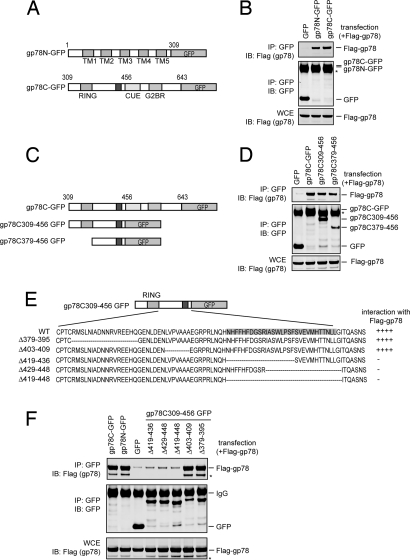
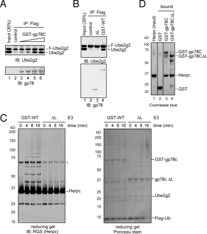
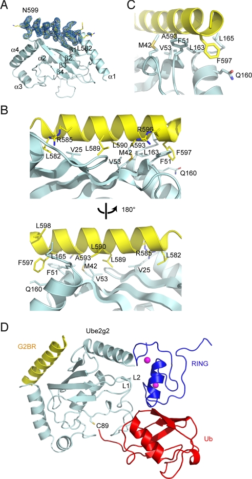
Lys-48-linked polyubiquitination regulates a variety of cellular processes by targeting ubiquitinated proteins to the proteasome for degradation. Although polyubiquitination had been presumed to occur by transferring ubiquitin molecules, one at a time, from an E2 active site to a substrate, we recently showed that the endoplasmic reticulum-associated RING finger ubiquitin ligase gp78 can mediate the preassembly of Lys-48-linked polyubiquitin chains on the catalytic cysteine of its cognate E2 Ube2g2 and subsequent transfer to a substrate. Active site-linked polyubiquitin chains are detected in cells on Ube2g2 and its yeast homolog Ubc7p, but how these chains are assembled is unclear. Here, we show that gp78 forms an oligomer via 2 oligomerization sites, one of which is a hydrophobic segment located in the gp78 cytosolic domain. We further demonstrate that a gp78 oligomer can simultaneously associate with multiple Ube2g2 molecules. This interaction is mediated by a novel Ube2g2 surface distinct from the predicted RING binding site. Our data suggest that a large gp78-Ube2g2 heterooligomer brings multiple Ube2g2 molecules into close proximity, allowing ubiquitin moieties to be transferred between neighboring Ube2g2s to form active site-linked polyubiquitin chains.


Conflict of interest statement

The authors declare no conflict of interest.

Figures

Fig. 1.
Fig. 1.
Oligomerization of gp78. (A) Schematic representation of the gp78 variants tested in B. (B) Both the cytosolic domain and the transmembrane segments of gp78 can interact with full-length gp78. Detergent extracts of 293T cells transfected with the indicated plasmids were subjected to immunoprecipitation (IP) followed by immunoblotting (IB) with the indicated antibodies. Note that the expressed gp78 proteins comigrate with IgG (*). WCE, whole cell extract. (C–F) Mapping the region in the gp78 cytosolic domain that is necessary for its self-association. (C) Schematic representation of the gp78 variants tested in D. (D) As in B, except that plasmids expressing the indicated gp78 variants were analyzed. (E) Schematic representation of the gp78 variants used in F. (F) As in B, except that plasmids expressing the indicated gp78 variants were analyzed. * indicates a gp78 degradation product.
Fig. 2.
Fig. 2.
Oligomerization of gp78C is required for Ube2g2 active site-linked polyubiquitination, but is dispensable for ubiquitin ligation activity. (A) Purified recombinant gp78C variants. (B) Polyubiquitinating activity of gp78CΔL. Polyubiquitination reaction conducted with E1, Ube2g2, Flag-Ub, and the indicated E3 ligases was analyzed under both nonreducing (−DTT) and reducing (+DTT) conditions by immunoblotting. Arrows indicate the mobility shift for Ube2g2 active site-linked ubiquitin chains upon treatment with DTT. * indicates Ube2g2∼Ub thioester. Note that the ubiquitin chains synthesized by gp78CΔL (ΔL) exhibit a pattern different from that generated by GST-gp78C (GST-WT) on a nonreducing gel. (C) gp78CΔL cannot synthesize ubiquitin chains on the Ube2g2 active site. Ubiquitin chains synthesized in the absence (no E3) or presence of the indicated gp78C variants were analyzed by immunoblotting. E2∼Ub(n), E2-linked ubiquitin chains. Note that the Flag antibody has a higher affinity for polyubiquitin chains than for monoubiquitin. (D) Purified recombinant MBP-gp78C variants. (E) Polyubiquitinating activity of MBP-gp78CΔL. Polyubiquitination reactions conducted with the indicated MBP-tagged gp78C variants were analyzed under both nonreducing (−DTT) and reducing (+DTT) conditions. Arrows indicate the mobility shift for Ube2g2 active site-linked ubiquitin chains upon treatment with DTT. (F) Ubiquitin chains synthesized by MBP-gp78CΔL (MBP-ΔL) are not linked to Ube2g2.
Fig. 3.
Fig. 3.
gp78CΔL stimulates the formation of free diubiquitin but is defective in building Ube2g2-linked diubiquitin. (A) As indicated by the experimental scheme, the transfer of Flag-UbK48R from Ube2g2 to an Ube2g2∼ubiquitin thioester was monitored by immunoblotting with Ube2g2 (E2) and Flag antibodies. * indicates a nonspecific band. (B) As indicated by the experimental scheme, the transfer of Flag-UbK48R from Ube2g2 to free ubiquitin was monitored by immunoblotting with Ube2g2 and Flag antibodies.
Fig. 4.
Fig. 4.
The monomeric gp78C mutant cannot promote Ube2g2 self-assembly and is defective in polyubiquitination of Herpc. (A) gp78C promotes Ube2g2 self-assembly. Equal amount of Flag-tagged (F-Ube2g2) and untagged Ube2g2 were mixed in the presence of either GST (control) or increasing amounts of GST-gp78C (lanes 3–6). Flag-Ube2g2 and its associated proteins were immunoprecipitated by anti-Flag agarose beads and analyzed by immunoblotting. Lane 1 shows input (30% of Ube2g2 used in the binding experiment). (B) gp78CΔL does not facilitate Ube2g2 self-assembly. As in A, except that both GST-gp78C (GST-WT) and gp78CΔL (ΔL) were tested at a single concentration. (C) gp78CΔL is defective in Herpc polyubiquitination. Polyubiquitination reactions conducted with E1, Ube2g2, Ub, Herpc, and the indicated gp78 variants were analyzed under reducing condition by immunoblotting and Ponceau staining. * indicates a nonspecific band. (D) gp78CΔL can interact with Herpc. The indicated GST-fusion proteins were immobilized on glutathione beads and incubated with purified Herpc (lanes 2–4). Bound proteins were eluted and analyzed by SDS/PAGE and Coomassie blue staining. Lane 1 shows Herpc input (30%).
Fig. 5.
Fig. 5.
The structure of Ube2g2 bound to the gp78 G2BR domain. (A) Overall structure showing the Ube2g2–G2BR complex. Contoured at 3.5 σ around G2BR is a 2FoFc σA-weighted annealed omit map omitting G2BR. (B) The contacts between G2BR and Ube2g2. (C) A close-up view on the most critical contacts around A593 and F597 of G2BR. (D) The geometry of G2BR binding compared with RING binding and the active site. RING domain (blue) of the c-Cbl–UbcH7 complex (Protein Data Bank ID code 1FBV), and Ub (red) of the Mms2-Ubc13∼Ub covalent complex (Protein Data Bank ID code 2GMI) are docked onto the Ube2g2–G2BR complex based on E2 structural alignments. Overlay matrices were determined by DALI.
Fig. 6.
Fig. 6.
Ube2g2–G2BR interaction is required for active site-linked polyubiquitination and substrate ubiquitination. (A) Interaction of Ube2g2 with gp78C is mediated by the G2BR domain. GST-tagged gp78C immobilized on glutathione beads was incubated with the indicated Ube2g2 variants. The bound proteins were analyzed by immunoblotting. (B) The Ube2g2–G2BR binding is required for Ube2g2 self-interaction. As in Fig. 4A except that the indicated untagged Ube2g2 variants were incubated with Flag–Ube2g2 in the absence (−) or presence (+) of GST-gp78C. (C and D) Surface plasmon resonance analyses of GST–gp78C–Ube2g2 interaction. (C) Representative sensorgrams for the binding of Ube2g2 to GST-gp78C. (D) The relative binding of wild-type Ube2g2 (E2 WT) and the quadruple mutant (E2 quad.) to GST-gp78C. (E) Active site-linked polyubiquitination by Ube2g2 variants. The reactions were analyzed under a nonreducing condition by immunoblotting. (F) Polyubiquitination of Herpc by Ube2g2 variants. As in E, except that the reaction also contains Herpc and that the reaction was analyzed under reducing condition.

References

    1. Weissman AM. Themes and variations on ubiquitylation. Nat Rev Mol Cell Biol. 2001;2:169–178. - PubMed
    1. Welchman RL, Gordon C, Mayer RJ. Ubiquitin and ubiquitin-like proteins as multifunctional signals. Nat Rev Mol Cell Biol. 2005;6:599–609. - PubMed
    1. Mukhopadhyay D, Riezman H. Proteasome-independent functions of ubiquitin in endocytosis and signaling. Science. 2007;315:201–205. - PubMed
    1. Pickart CM. Mechanisms underlying ubiquitination. Annu Rev Biochem. 2001;70:503–533. - PubMed
    1. Jin J, Li X, Gygi SP, Harper JW. Dual E1 activation systems for ubiquitin differentially regulate E2 enzyme charging. Nature. 2007;447:1135–1138. - PubMed

Publication types

MeSH terms

Associated data