Review
. 2009 May;35(3):528-48.
doi: 10.1093/schbul/sbn187. Epub 2009 Feb 17.

The neurodevelopmental hypothesis of schizophrenia, revisited

Affiliations
Review

The neurodevelopmental hypothesis of schizophrenia, revisited

S Hossein Fatemi et al. Schizophr Bull. 2009 May.

Abstract

While multiple theories have been put forth regarding the origin of schizophrenia, by far the vast majority of evidence points to the neurodevelopmental model in which developmental insults as early as late first or early second trimester lead to the activation of pathologic neural circuits during adolescence or young adulthood leading to the emergence of positive or negative symptoms. In this report, we examine the evidence from brain pathology (enlargement of the cerebroventricular system, changes in gray and white matters, and abnormal laminar organization), genetics (changes in the normal expression of proteins that are involved in early migration of neurons and glia, cell proliferation, axonal outgrowth, synaptogenesis, and apoptosis), environmental factors (increased frequency of obstetric complications and increased rates of schizophrenic births due to prenatal viral or bacterial infections), and gene-environmental interactions (a disproportionate number of schizophrenia candidate genes are regulated by hypoxia, microdeletions and microduplications, the overrepresentation of pathogen-related genes among schizophrenia candidate genes) in support of the neurodevelopmental model. We relate the neurodevelopmental model to a number of findings about schizophrenia. Finally, we also examine alternate explanations of the origin of schizophrenia including the neurodegenerative model.


Figures

Fig. 1.
Fig. 1.
Magnetic Resonance Imaging Reveals Significant (P < .05) Brain Atrophy in Multiple Brain Areas of the 35-d-Old Virally Infected Mouse Offspring (Right Panel) as Compared With Sham-Infected Mice (Left Panel). Originally published in Fatemi et al.
Fig. 2.
Fig. 2.
A Hypothesis of How Prenatal Viral Infection Could Contribute to the Development of Schizophrenia. Prenatal viral infection may lead to (1) activation of DNA methyltransferase 1 (DNMT1) that in turn changes methylation of promoters for a variety of genes leading to altered levels of molecules such as glutamic acid decarboxylase 67-kDa protein (GAD67) and reelin (S.H. Fatemi, unpublished observations)., These changes may result in abnormal development and altered γ-aminobutyric acid (GABA) signaling and subsequent genesis of schizophrenia; (2) activation of the maternal immune response leading to altered levels of cytokines including interleukin (IL)-1β, IL-6, and tumor necrosis factor α (TNF-α),, that regulate normal brain development.– Changes may lead to abnormal cortical development and, ultimately, schizophrenia; and (3) altered expression of genes that are involved in cell-cell communication and changes in cell structure due to chronic actin depolymerization (S.H. Fatemi and M. Peoples, unpublished observations, 2007) may lead to dysregulation of multiple signaling systems that have been observed in schizophrenia. *, Pathways that require more substantial support.
Fig. 3.
Fig. 3.
Reelin is Reduced in Hippocampus of Individuals With Schizophrenia and in Cerebral Cortex Following Prenatal Viral Infection. A. The values expressed on the y-axis are reelin-positive adjusted cell densities per square millimeter localized to hippocampal CA4 areas in control and schizophrenic subjects. The number of brains used is 15 (control) and 15 (schizophrenic). Each point is the mean for 2–4 sections analyzed per brain. A crossbar localized over each scatter plot represents mean Reelin-positive adjusted cell density value per group. Mean values for schizophrenic subjects are significantly reduced when compared with control values (analysis of variance, P < .05). B. The top panel shows a graph depicting the hemispheric Reelin-positive Cajal-Retzius (CR) cell counts in layer I of the cortex of prenatally infected (I) and sham-infected control (C) animals. The number of Reelin-positive CR cells was significantly reduced in infected brains compared with control brains (P < .0001). The lower panel shows light micrographs of layer I–II in coronal sections of prenatally infected and sham-infected cortex. Originally published in Fatemi et al.,
Fig. 4.
Fig. 4.
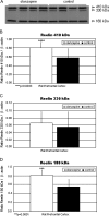
(A) Reelin Bands of 410, 330, and 180 kDa From the Prefrontal Cortex Homogenates (70 μg protein per lane) of Representative Olanzapine-Treated and Control Rats Are Shown. Mean Reelin 410 (B), 330 (C), and 180 (D) kDa/β-actin ratios for olanzapine-treated (filled histogram bars) and control rats (unfilled histogram bars) are shown. Levels of Reelin 410 kDa/β-actin (B) and Reelin 180 kDa/β-actin (D) were significantly increased vs controls (P = .0033 and .0001, respectively). Reelin 330 kDa/β-actin (C) was nonsignificantly increased vs controls. Origianlly published in Fatemi et al.
Fig. 5.
Fig. 5.
Effects of Psychotropic Agents on Catechol-O-Methyltransferase (COMT) Expression in Rat Frontal Cortex. A, C, E, G, I, and K correspond to protein levels from frontal cortices of clozapine-, fluoxetine-, haloperidol-, lithium-, olanzapine-, and sodium valproate–treated rat brains, respectively. B, D, F, H, J, and L correspond to protein levels from frontal cortices of saline-treated rat brains, respectively. Originally published in Fatemi and Folsom.

Similar articles

Cited by

References

    1. Rapoport JL, Addington AM, Frangou S, Psych MR. The neurodevelopmental model of schizophrenia: update 2005. Mol Psychiatry. 2005;10:434–449. - PubMed
    1. Fatemi SH. Prenatal viral infection, brain development and schizophrenia. In: Fatemi SH, editor. Neuropsychiatric Disorders and Infection. London, UK: Taylor and Francis; 2005.
    1. Krapelin E. Psychiatrie. 4th ed. Ein lehrbuch für studirende und ärzte [Psychiatry 4th Ed: A Textbook for Students and Physicians] Leipzeig, Germany: Abel; 1893.
    1. Brown AS, Begg MD, Gravenstein S, et al. Serologic evidence of prenatal influenza in the etiology of schizophrenia. Arch Gen Psychiatry. 2004;61:774–780. - PubMed
    1. Fatemi SH. Schizophrenia. In: Fatemi SH, Clayton PJ, editors. Medical Basis of Psychiatry. New York, NY: Humana Press; 2008. pp. 85–108.

Publication types

MeSH terms