

Dose perturbations from implanted helical gold markers in proton therapy of prostate cancer
- PMID: 19223836
- PMCID: PMC2949274
- DOI: 10.1120/jacmp.v10i1.2875
Dose perturbations from implanted helical gold markers in proton therapy of prostate cancer
Abstract
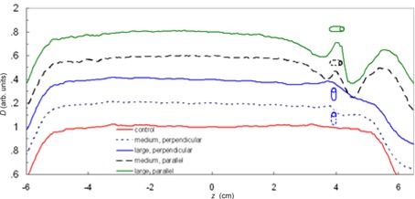
Implanted gold fiducial markers are widely used in radiation therapy to improve targeting accuracy. Recent investigations have revealed that metallic fiducial markers can cause severe perturbations in dose distributions for proton therapy, suggesting smaller markers should be considered. The objective of this study was to estimate the dosimetric impact of small gold markers in patients receiving proton therapy for prostate cancer. Small, medium, and large helical wire markers with lengths of 10 mm and helix diameters of 0.35 mm, 0.75 mm, and 1.15 mm, respectively, were implanted in an anthropomorphic phantom. Radiographic visibility was confirmed using a kilovoltage x-ray imaging system, and dose perturbations were predicted from Monte Carlo simulations and confirmed by measurements. Monte Carlo simulations indicated that size of dose perturbation depended on marker size, orientation, and distance from the beam's end of range. Specifically, the perturbation of proton dose for the lateral-opposed-pair treatment technique was 31% for large markers and 23% for medium markers in a typical oblique orientation. Results for perpendicular and parallel orientations were respectively lower and higher. Consequently, these markers are not well suited for use in patients receiving proton therapy for prostate cancer. Dose perturbation was not observed for the small markers, but these markers were deemed too fragile for transrectal implantation in the prostate.
Figures

References
-
- Sorcini B, Tilikidis A. Clinical application of image‐guided radiotherapy, IGRT (on the Varian OBI platform). Cancer Radiother. 2006;10(5):252–57. - PubMed
-
- Sorensen SP, Chow PE, Kriminski S, Medin PM, Solberg TD. Image‐guided radiotherapy using a mobile kilovoltage x‐ray device. Med Dosim. 2006;31(1): 40–50. - PubMed
-
- Huntzinger C, Munro P, Johnson S, et al. Dynamic targeting image‐guided radiotherapy. Med Dosim. 2006;31(2):113–25. - PubMed
-
- Letourneau D, Martinez AA, Lockman D, et al. Assessment of residual error for online cone‐beam CT‐guided treatment of prostate cancer patients. Int J Radiat Oncol Biol Phys. 2005;62(4):1239–46. - PubMed
-
- Gall KP, Verhey LJ, Wagner M. Computer‐assisted positioning of radiotherapy patients using implanted radiopaque fiducials. Med Phys. 1993;20(4):1153–59. - PubMed
Publication types
MeSH terms
Substances
Grants and funding
LinkOut - more resources
Full Text Sources
Other Literature Sources
Medical
