

Automated quality assurance for image-guided radiation therapy
- PMID: 19223842
- PMCID: PMC5720496
- DOI: 10.1120/jacmp.v10i1.2919
Automated quality assurance for image-guided radiation therapy
Abstract
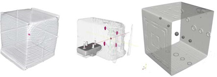
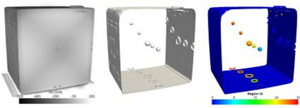
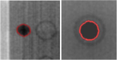
The use of image-guided patient positioning requires fast and reliable Quality Assurance (QA) methods to ensure the megavoltage (MV) treatment beam coincides with the integrated kilovoltage (kV) or volumetric cone-beam CT (CBCT) imaging and guidance systems. Current QA protocol is based on visually observing deviations of certain features in acquired kV in-room treatment images such as markers, distances, or HU values from phantom specifications. This is a time-consuming and subjective task because these features are identified by human operators. The method implemented in this study automated an IGRT QA protocol by using specific image processing algorithms that rigorously detected phantom features and performed all measurements involved in a classical QA protocol. The algorithm was tested on four different IGRT QA phantoms. Image analysis algorithms were able to detect QA features with the same accuracy as the manual approach but significantly faster. All described tests were performed in a single procedure, with acquisition of the images taking approximately 5 minutes, and the automated software analysis taking less than 1 minute. The study showed that the automated image analysis based procedure may be used as a daily QA procedure because it is completely automated and uses a single phantom setup.
Figures


Similar articles
-
Efficient quality assurance method with automated data acquisition of a single phantom setup to determine radiation and imaging isocenter congruence.J Appl Clin Med Phys. 2019 Oct;20(10):127-133. doi: 10.1002/acm2.12723. Epub 2019 Sep 19. J Appl Clin Med Phys. 2019. PMID: 31535781 Free PMC article.
-
Initial application of a geometric QA tool for integrated MV and kV imaging systems on three image guided radiotherapy systems.Med Phys. 2011 May;38(5):2335-41. doi: 10.1118/1.3570768. Med Phys. 2011. PMID: 21776767
-
A novel phantom and procedure providing submillimeter accuracy in daily QA tests of accelerators used for stereotactic radiosurgery*.J Appl Clin Med Phys. 2016 Jul 8;17(4):246-253. doi: 10.1120/jacmp.v17i4.6295. J Appl Clin Med Phys. 2016. PMID: 27455506 Free PMC article.
-
An overview of volumetric imaging technologies and their quality assurance for IGRT.Acta Oncol. 2008;47(7):1271-8. doi: 10.1080/02841860802244182. Acta Oncol. 2008. PMID: 18618343 Review.
-
A review of image-guided radiotherapy.Radiol Phys Technol. 2009 Jan;2(1):1-12. doi: 10.1007/s12194-008-0045-y. Epub 2008 Dec 16. Radiol Phys Technol. 2009. PMID: 20821123 Review.
Cited by
-
Isocenter verification for linac-based stereotactic radiation therapy: review of principles and techniques.J Appl Clin Med Phys. 2011 Nov 15;12(4):3645. doi: 10.1120/jacmp.v12i4.3645. J Appl Clin Med Phys. 2011. PMID: 22089022 Free PMC article.
-
Semi-automated IGRT QA using a cone-shaped scintillator screen detector for proton pencil beam scanning treatments.Phys Med Biol. 2019 Apr 5;64(8):085004. doi: 10.1088/1361-6560/ab056d. Phys Med Biol. 2019. PMID: 30736026 Free PMC article.
-
An AI-based universal phantom analysis method based on XML-SVG wireframes with novel functional object identifiers.Phys Med Biol. 2023 Jul 14;68(14):10.1088/1361-6560/acdb44. doi: 10.1088/1361-6560/acdb44. Phys Med Biol. 2023. PMID: 37267994 Free PMC article.
-
Efficient quality assurance method with automated data acquisition of a single phantom setup to determine radiation and imaging isocenter congruence.J Appl Clin Med Phys. 2019 Oct;20(10):127-133. doi: 10.1002/acm2.12723. Epub 2019 Sep 19. J Appl Clin Med Phys. 2019. PMID: 31535781 Free PMC article.
-
Automating quality assurance of digital linear accelerators using a radioluminescent phosphor coated phantom and optical imaging.Phys Med Biol. 2016 Sep 7;61(17):L29-37. doi: 10.1088/0031-9155/61/17/L29. Epub 2016 Aug 12. Phys Med Biol. 2016. PMID: 27514654 Free PMC article.
References
-
- Saw CB, Yang Y, Li F, et al. Performance characteristics and quality assurance aspects of kilovoltage cone‐beam CT on medical linear accelerator. Med Dosim. 2007;32(2):80–85. - PubMed
-
- Bissonnette J‐P. Quality assurance of image‐guidance technologies. Semin Radiat Oncol. 2007;17(4):278–86. - PubMed
-
- Yoo S, Kim GY, Hammoud R, et al. A quality assurance program for the on‐board imagers. Med Phys. 2006;33(11):4431–47. - PubMed
-
- Sharpe MB, Moseley DJ, Purdie TG, Islam M, Siewerdsen JH, Jaffray DA. The stability of mechanical calibration for a kV cone beam computed tomography system integrated with linear accelerator. Med Phys. 2006;33(1):136–44. - PubMed
MeSH terms
LinkOut - more resources
Full Text Sources
Other Literature Sources
Medical
