. 2009 Apr;17(4):675-84.
doi: 10.1038/mt.2008.307. Epub 2009 Feb 17.

Redundant and synergistic mechanisms control the sequestration of blood-born adenovirus in the liver

Affiliations

Redundant and synergistic mechanisms control the sequestration of blood-born adenovirus in the liver

Nelson C Di Paolo et al. Mol Ther. 2009 Apr.

Abstract

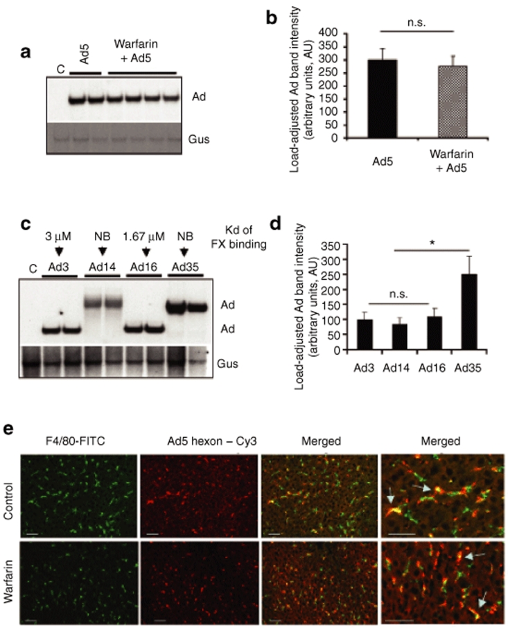
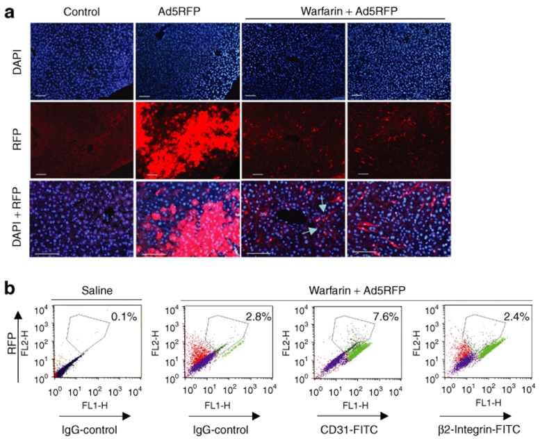
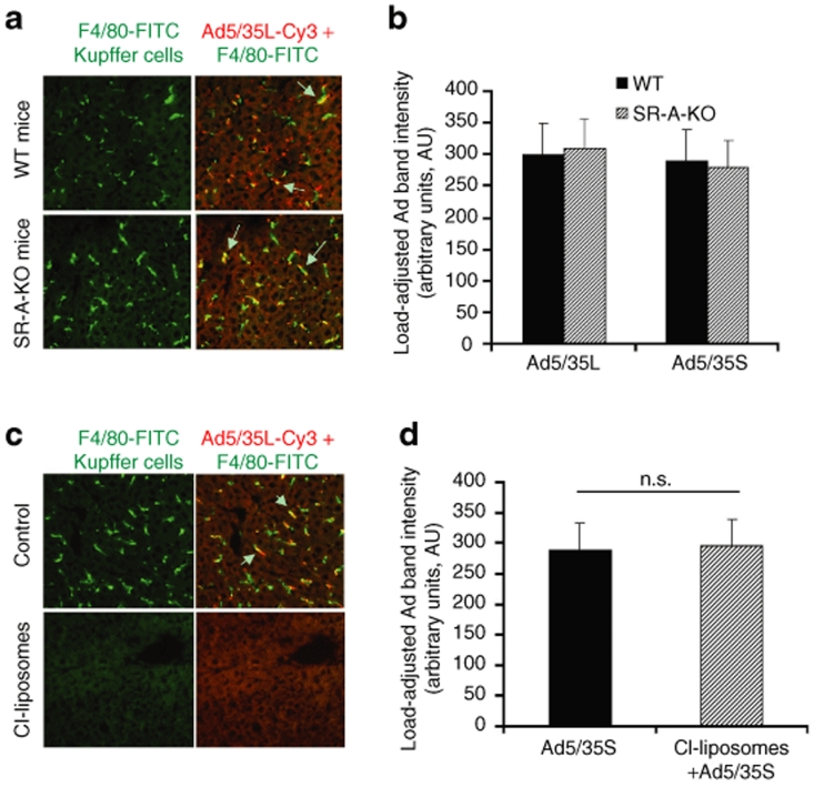
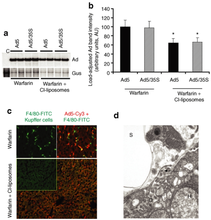
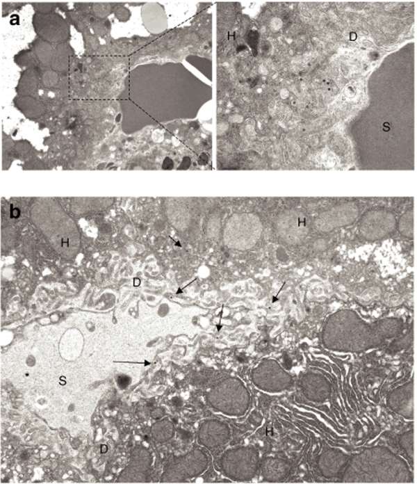
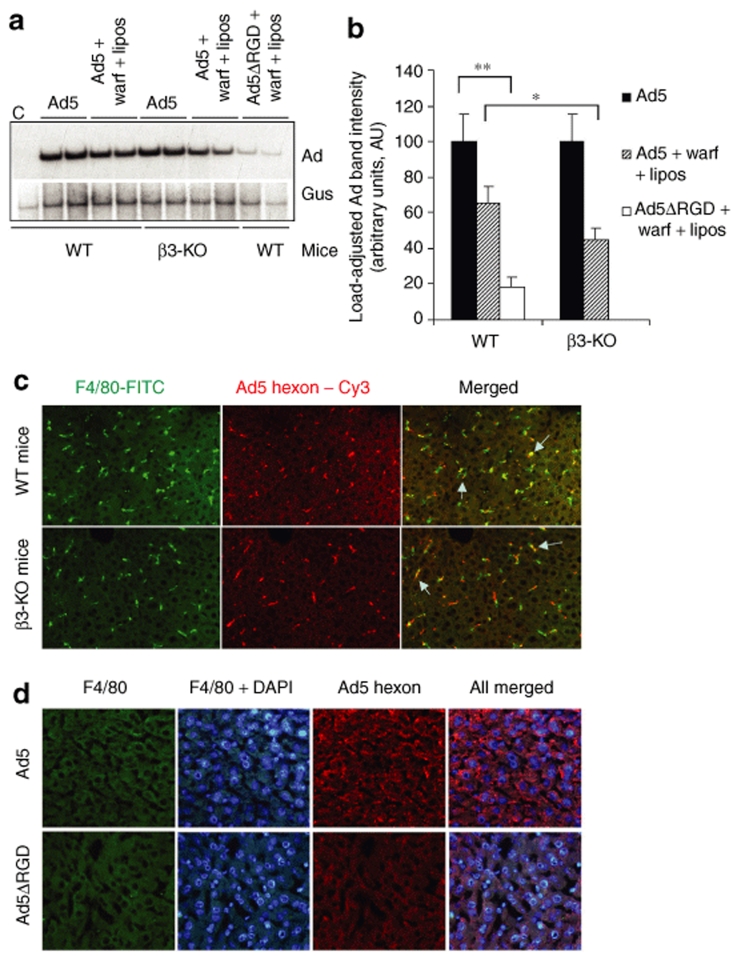
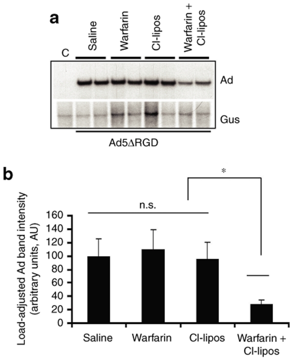
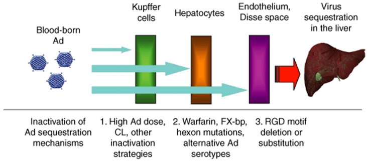
Human adenovirus (Ad) is a ubiquitous pathogen causing a wide range of diseases. Although the interactions of human Ad serotype 5 (Ad5) with susceptible cells in vitro are known in great detail, host factors controlling the tissue specificity of Ad5 infection in vivo remain poorly understood. Here, we analyzed the mechanisms of sequestration by the liver for blood-born human Ads and Ad5-based vectors. Our data suggest that several known mechanisms that lead to Ad5 sequestration by the liver become engaged in a redundant, sequential, and synergistic manner to ensure the rapid clearance of circulating virus particles from the blood. These mechanisms include (i) trapping of the virus by liver residential macrophages, Kupffer cells; (ii) Ad5 hepatocyte infection via blood factor-hexon interactions; and (iii) Ad5 penton RGD motif-mediated interactions with liver endothelial cells and hepatocytes, mediating virus retention in the space of Disse. More important, we show that when all of these mechanisms are simultaneously inactivated via mutations of Ad5 capsid proteins and pharmacological interventions, virus sequestration by the liver is markedly reduced. Therefore, our study is the first demonstration of the principal possibility of ablating the sequestration of blood-born Ad in the liver via specific inactivation of a defined set of mechanisms that control this process.


Figures

<b>Figure 1</b>
Figure 1
Sequestration of blood-born adenovirus (Ad) in the liver tissue after intravenous virus injection occurs independently of virus binding to blood coagulation factors. (a) Ad5 vector was injected into mice mock treated (Ad5 lanes) or pretreated with warfarin (warfarin lanes), and livers were harvested 1 hour after virus injection and processed for Southern blotting as described in Materials and Methods. Each lane represents liver samples harvested from a mouse individually injected with Ad (biological replicates). (b) Quantitative representation of vector accumulation in livers determined by PhosphorImager analysis of Ad-specific bands shown in a after adjustment of Ad DNA signal intensities for the Gus gene signal intensities for corresponding vectors using ImageQuant software. N = 4. n.s.—not statistically significant. (c) Southern blot analysis of genomes of wild-type human Ad present in the liver 1 hour after intravenous virus injection. Affinity of factor X (FX) binding for indicated serotypes is shown. Gus—mouse β-glucuronidase gene. C—Control liver DNA from a mouse injected with virus dilution buffer (phosphate-buffered saline) only. NB—not binding. The affinity of Ad3, Ad16, and Ad35 for FX was previously reported in ref. 49. The lack of Ad14 binding to FX is a personal communication of A. Lieber (University of Washington, 20 November 2008). Conservative species B Ad–specific DNA probe corresponding to E4 region was used for detection of species B genomic DNA in hybridization reaction. (d) Quantitative representation of vector accumulation in livers determined by PhosphorImager analysis of Ad-specific bands shown in c after adjustment of Ad DNA signal intensities for the Gus gene signal intensities for corresponding vectors using ImageQuant software. N = 3. n.s.—not statistically significant. *P < 0.05. (e) Association of Ad vectors with Kupffer cells of mice treated with warfarin or mock-treated controls. Ads were injected into the tail vein of C57Bl/6 mice. One hour later, livers were recovered and immediately frozen in an optimal cutting temperature compound. To visualize Kupffer cells (KCs), fixed liver sections were stained with anti-F4/80 antibody conjugated with fluorescein isothiocyanate (FITC) (green). Ad particles were visualized after staining the liver sections with Cy3-labeled anti-hexon polyclonal antibodies (red). Images of representative fields were taken with red and green filters and were then superimposed to reveal Ad association with KCs (yellow, merged). The overlapping of Kupffer cell–specific staining and Ad-specific staining is indicated by arrows.
<b>Figure 2</b>
Figure 2
Inactivation of blood coagulation factors leads to adenovirus (Ad) transduction of liver sinusoid endothelial cells. (a) In vivo transduction of hepatocytes with Ad5RFP after systemic vector application in control, mock-treated, and warfarin-treated mice. Twenty-four hours after intravenous Ad injection, livers were recovered and serial sections of formalin-fixed tissues were prepared. To visualize red fluorescent protein (RFP) fluorescence, images of sections were taken under ultraviolet light. Representative fields are shown. Magnification, ×200 on the two left sets of panels and ×400 on the right sets of panels. Note that the treatment of mice with warfarin completely eliminates hepatocyte transduction. However, sinusoid endothelial cells express RFP (indicated by arrows) in warfarin-treated mice. Two representative fields are shown. N = 4. (b) Analysis of surface markers of RFP expressing cells in warfarin-treated mice using flow cytometry (N = 5, the average percentage of RFP-positive cells is shown on the dot plots). Note that the RFP expressing cells are stained positive with antibody for CD31, endothelial cell marker, and not with antibody specific for β2-integrin, which is the marker for hematopoietic cells. DAPI, 4′,6-diamidino-2-phenylindole; FITC, fluorescein isothiocyanate; IgG, immunoglobulin G.
<b>Figure 3</b>
Figure 3
Kupffer cell elimination does not prevent the sequestration of blood-born adenovirus (Ad) in the liver. (a) Association of Ad vectors with Kupffer cells of wild-type (WT) mice or mice knockout for scavenger receptor A (SR-A-KO) gene. One hour after intravenous virus injection, livers were recovered and immediately frozen in optimal cutting temperature compound. To visualize KCs, fixed liver sections were stained with anti-F4/80 antibody (green). Ad particles were visualized after staining liver sections with Cy3-labeled anti-hexon polyclonal antibodies (red). Images of representative fields were taken with red and green filters and were then superimposed to reveal Ad association with KCs (yellow). The overlapping of Kupffer cell–specific staining and Ad-specific staining is indicated by arrows. (b) Quantitative representation of vector accumulation in livers determined by Southern blot analysis followed by ImageQuant data processing of Ad-specific bands are shown in Supplementary Figure S2A after adjustment of Ad DNA signal intensities for the Gus gene signal intensities. N = 4. (c) Wild-type mice were treated with clodronate liposomes as described in Materials and Methods. Furthermore, 24 hours after the treatment of mice with clodronate liposomes and 1 hour after intravenous virus injection, livers were recovered and immediately frozen in optimal cutting temperature compound. To visualize KCs, fixed liver sections were stained with anti-F4/80 antibody (green). Note that the treatment of mice with clodronate liposomes completely eliminates Kupffer cells from the liver. (d) Quantitative representation of the Southern blot analysis of Ad vector genomes' association with livers of wild-type mice treated with clodronate liposomes 1 hour after intravenous virus injection. Following hybridizations, the membranes were exposed to PhophorImager screens and the intensity volumes of Ad-specific and Gus-specific bands were recorded and processed using ImageQuant software. N = 4. n.s.—not statistically significant.
<b>Figure 4</b>
Figure 4
The treatment of mice with warfarin and clodronate liposomes allows for a partial reduction of the levels of adenovirus (Ad) DNA sequestered by the liver after intravenous virus administration. (a) Southern blot analysis for Ad vector genomes associated with livers of wild-type mice 1 hour after intravenous virus injection. Mice were treated with warfarin only or with a combination of warfarin and clodronate liposomes. Duplicate samples for each group are shown. Gus—mouse β-glucuronidase gene. Control—liver DNA from a mouse injected with virus dilution buffer (phosphate-buffered saline) only. (b) Quantitative representation of Ad accumulation in livers determined by PhosphorImager analysis of Ad-specific bands shown in a after adjustment of Ad DNA signal intensities for the Gus gene signal intensities for corresponding vectors. *P < 0.05. (c) Distribution of Ad particles in the livers of mice treated with warfarin or with a combination of warfarin and clodronate liposomes 1 hour after intravenous virus injection. Liver sections were stained with anti-F4/80 antibody to detect Kupffer cells (green) and with anti-Ad hexon antibody to detect Ad particles (red). Note that the large number of Ad particles was colocalized with Kupffer cells in warfarin-treated animals, whereas Ad particles were associated with liver sinusoids in mice treated with both of drugs. (d) Ad particles (shown by the arrow) localize to a subendothelial Disse space in the livers of mice treated with both warfarin and clodronate liposomes. Electron microscopy analysis was done on ultrathin sections of livers harvested 1 hour after intravenous virus injection. D—Disse space; H—hepatocyte; S—liver sinusoidal space. Magnification ×21,000.
<b>Figure 5</b>
Figure 5
Visualization of adenovirus (Ad) distribution in liver tissue 1 hour after intravenous virus injection. (a) Ad particles present in hepatic sinusoids in a space of Disse; magnification of main image: ×4,400; enlarged image ×21,000. (b) Representative image showing distribution of free Ad particles in the Disse space (indicated by arrows). Magnification × 7,500. D—Disse space; H—hepatocyte; S—liver sinusoidal space.
<b>Figure 6</b>
Figure 6
Adenovirus (Ad) penton RGD motifs play a critical role in supporting the sequestration of the blood-born Ad in the liver. (a) Wild-type (WT) or β3-integrin knockout mice (β3-KO) were treated with a combination of warfarin and liposomes and injected intravenously with Ad5 vector. In addition to drug treatment, WT mice were also injected with Ad5ΔRGD vector. One hour after virus injection, livers were harvested, and total liver DNA was purified and then subjected to a Southern blot analysis as described in Figure 1. Duplicate samples for each group are shown. Gus—mouse β-glucuronidase gene. C—Control liver DNA from a mouse injected with virus dilution buffer (phosphate-buffered saline) only. (b) Quantitative representation of Ad accumulation in livers determined by PhosphorImager analysis of Ad-specific bands shown in a after adjustment of Ad DNA signal intensities for the Gus gene signal intensities for corresponding vectors. *P < 0.05. **P < 0.01. (c) Distribution of Ad particles in the livers of wild-type or β3-KO mice 1 hour after intravenous virus injection. Liver sections were stained with anti-F4/80 antibody to detect Kupffer cells (green) and with anti-Ad hexon antibody to detect Ad particles (red). Colocalization of Ad-specific staining with Kupffer cell staining appears in yellow (merged) and is indicated by arrows. (d) Distribution of Ad and Ad5ΔRGD particles in the livers of mice treated with a combination of warfarin and clodronate liposomes 1 hour after intravenous injection. Liver sections were stained with anti-F4/80 antibody to ensure complete elimination of Kupffer cells with clodronate liposomes (green) as well as with 4′,6-diamidino-2-phenylindole (DAPI) (blue) and anti-Ad hexon antibody to detect Ad particles (red).
<b>Figure 7</b>
Figure 7
Sequestration of blood-born Ad5ΔRGD vector in the liver is reduced only in mice treated with both warfarin and clodronate liposomes. (a) Wild-type mice were individually treated with a saline, warfarin, clodronate liposomes, or a combination of warfarin and liposomes and injected intravenously with Ad5ΔRGD vector. One hour after virus injection, livers were harvested, and total liver DNA was purified and subjected to Southern blot analysis as described in Figure 1. Duplicate samples for each group are shown. Gus—mouse β-glucuronidase gene. C—Control liver DNA from a mouse injected with virus dilution buffer (phosphate-buffered saline) only. (b) Quantitative representation of Ad5ΔRGD accumulation in livers determined by PhosphorImager analysis of adenovirus (Ad)-specific bands shown in a after adjustment of Ad DNA signal intensities for the Gus gene signal intensities for corresponding vectors. *P < 0.05. n.s.—not statistically significant.
<b>Figure 8</b>
Figure 8
Schematic representation of mechanisms mediating the sequestration of blood-born adenovirus (Ad) in the liver and approaches to inactivate them. Upon entry of Ad into a bloodstream, a defined set of specific molecular mechanisms become engaged in a redundant, synergistic, and orderly manner to ensure the clearance of blood-born Ad from circulation. If small amounts of Ad particles appear in blood, the virus trapping by Kupffer cells works as a first dominant mechanism, mediating Ad sequestration in the liver. When the Ad dose exceeds the capacity of Kupffer cells to trap the virus, hepatocytes absorb blood-born Ad particles in a blood factor–dependent manner, serving as a second dominant mechanism mediating sequestration of blood-born Ad. However, when the Ad dose is high and both the Kupffer cells and blood factor pathways are inactivated, sinusoid endothelial cells and the anatomical architecture of liver sinusoids become the third line of defense that sequesters Ad particles in an RGD motif–dependent manner. CL—clodronate liposomes; FX-bp—FX binding protein.

References

    1. Thomas CE, Ehrhardt A., and , Kay MA. Progress and problems with the use of viral vectors for gene therapy. Nat Rev Gen. 2003;4:346–358. - PubMed
    1. Alemany R, Suzuki K., and , Curiel DT. Blood clearance rates of adenovirus type 5 in mice. J Gen Virol. 2000;81:2605–2609. - PubMed
    1. Worgall S, Wolff G, Falck-Pedersen E., and , Crystal RG. Innate immune mechanisms dominate elimination of adenoviral vectors following in vivo administration. Hum Gene Ther. 1997;8:37–44. - PubMed
    1. Bergelson JM, Cunningham JA, Droguett G, Kurt-Jones EA, Krithivas A, Hong JS, et al. Isolation of a common receptor for Coxsackie B viruses and adenoviruses 2 and 5. Science. 1997;275:1320–1323. - PubMed
    1. Gaggar A, Shayakhmetov D., and , Lieber A. CD46 is a cellular receptor for group B adenoviruses. Nat Med. 2003;9:1408–1412. - PubMed

Publication types

LinkOut - more resources