

Taurine: a potential marker of apoptosis in gliomas
- PMID: 19223899
- PMCID: PMC2653765
- DOI: 10.1038/sj.bjc.6604933
Taurine: a potential marker of apoptosis in gliomas
Abstract
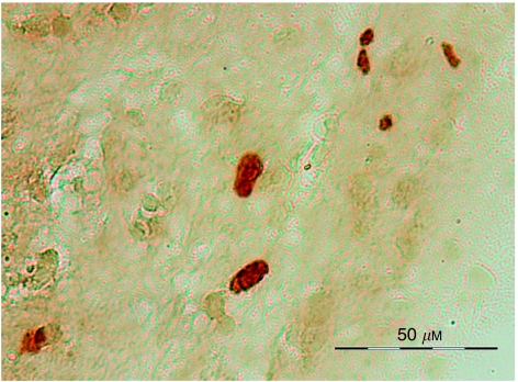
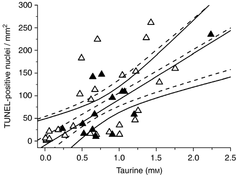
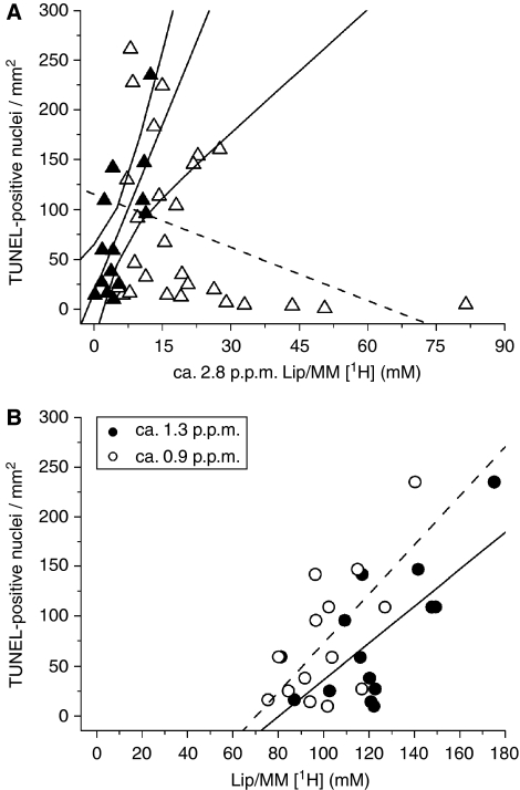
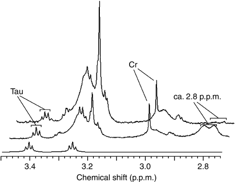
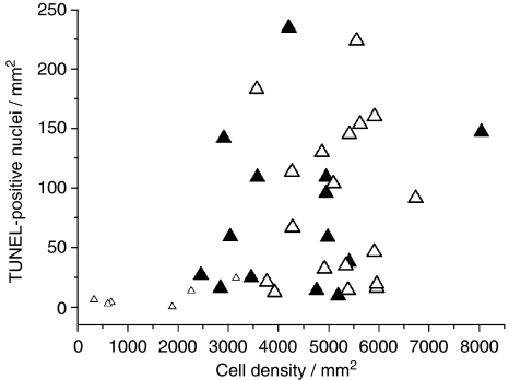
New cancer therapies are being developed that trigger tumour apoptosis and an in vivo method of apoptotic detection and early treatment response would be of great value. Magnetic resonance spectroscopy (MRS) can determine the tumour biochemical profile in vivo, and we have investigated whether a specific spectroscopic signature exists for apoptosis in human astrocytomas. High-resolution magic angle spinning (HRMAS) (1)H MRS provided detailed (1)H spectra of brain tumour biopsies for direct correlation with histopathology. Metabolites, mobile lipids and macromolecules were quantified from presaturation HRMAS (1)H spectra acquired from 41 biopsies of grades II (n=8), III (n=3) and IV (n=30) astrocytomas. Subsequently, TUNEL and H&E staining provided quantification of apoptosis, cell density and necrosis. Taurine was found to significantly correlate with apoptotic cell density (TUNEL) in both non-necrotic (R=0.727, P=0.003) and necrotic (R=0.626, P=0.0005) biopsies. However, the ca 2.8 p.p.m. polyunsaturated fatty acid peak, observed in other studies as a marker of apoptosis, correlated only in non-necrotic biopsies (R=0.705, P<0.005). We suggest that the taurine (1)H MRS signal in astrocytomas may be a robust apoptotic biomarker that is independent of tumour necrotic status.
Figures
References
-
- Cheng LL, Chang IW, Louis DN, Gonzalez RG (1998) Correlation of high-resolution magic angle spinning proton magnetic resonance spectroscopy with histopathology of intact human brain tumor specimens. Cancer Res 58: 1825–1832 - PubMed
-
- Delikatny EJ, Cooper WA, Brammah S, Sathasivam N, Rideout DC (2002) Nuclear magnetic resonance-visible lipids induced by cationic lipophilic chemotherapeutic agents are accompanied by increased lipid droplet formation and damaged mitochondria. Cancer Res 62: 1394–1400 - PubMed
-
- Di Vito M, Lenti L, Knijn A, Iorio E, D'Agostino F, Molinari A, Calcabrini A, Stringaro A, Meschini S, Arancia G, Bozzi A, Strom R, Podo F (2001) H NMR-visible mobile lipid domains correlate with cytoplasmic lipid bodies in apoptotic T-lymphoblastoid cells. Biochim Biophys Acta 1530: 47–66 - PubMed
Publication types
MeSH terms
Substances
Grants and funding
LinkOut - more resources
Full Text Sources
Medical
