. 2009;4(2):e4503.
doi: 10.1371/journal.pone.0004503. Epub 2009 Feb 18.

Cellular expression, trafficking, and function of two isoforms of human ULBP5/RAET1G

Affiliations

Cellular expression, trafficking, and function of two isoforms of human ULBP5/RAET1G

Robert A Eagle et al. PLoS One. 2009.

Erratum in

Abstract

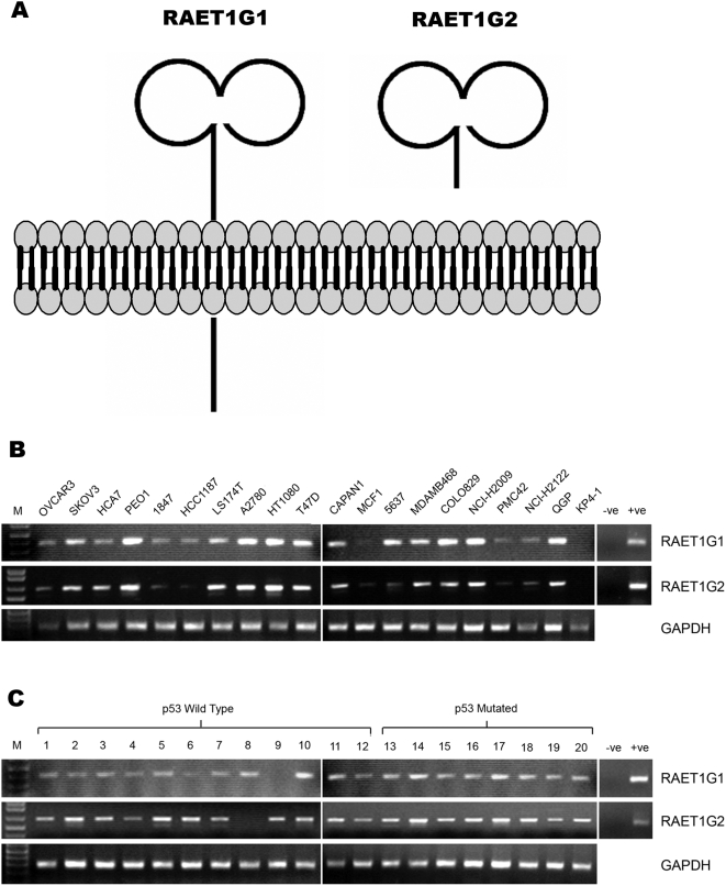
Background: The activating immunoreceptor NKG2D is expressed on Natural Killer (NK) cells and subsets of T cells. NKG2D contributes to anti-tumour and anti-viral immune responses in vitro and in vivo. The ligands for NKG2D in humans are diverse proteins of the MIC and ULBP/RAET families that are upregulated on the surface of virally infected cells and tumours. Two splicing variants of ULBP5/RAET1G have been cloned previously, but not extensively characterised.

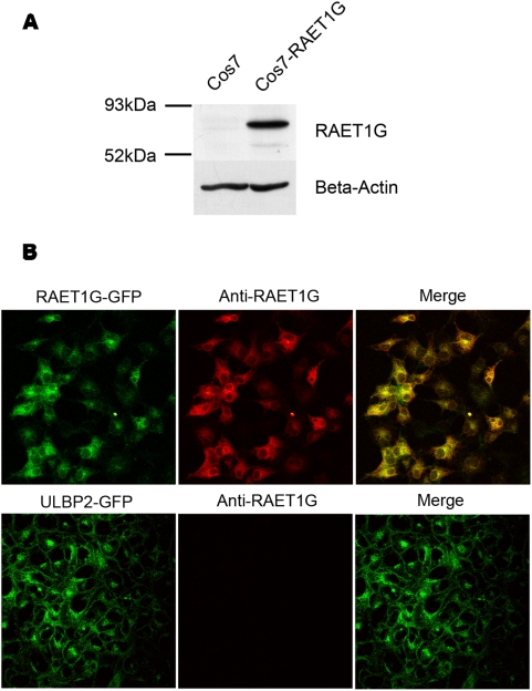
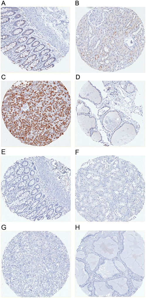
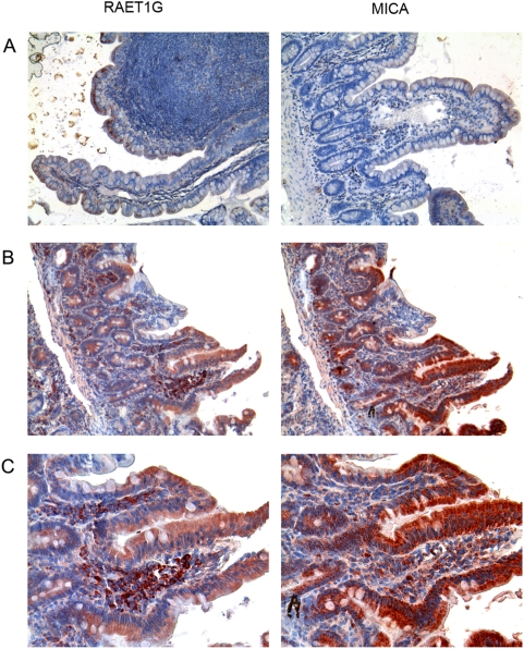
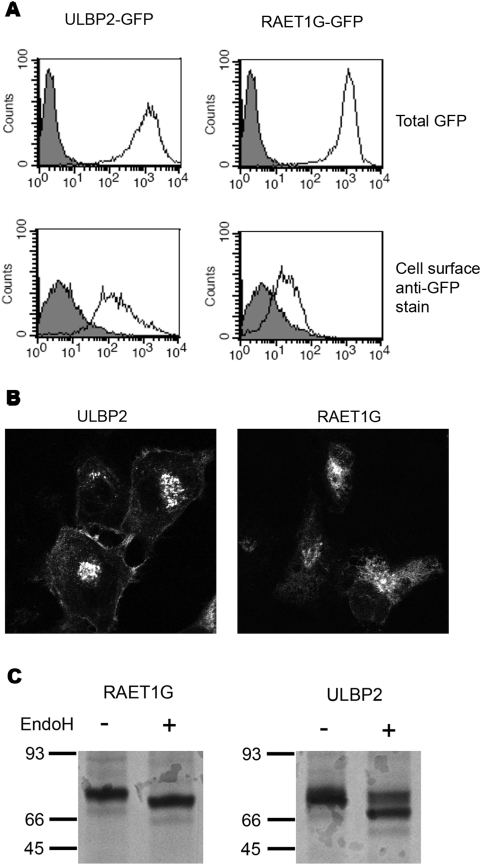
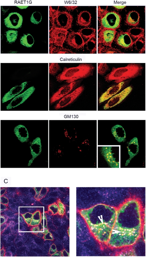
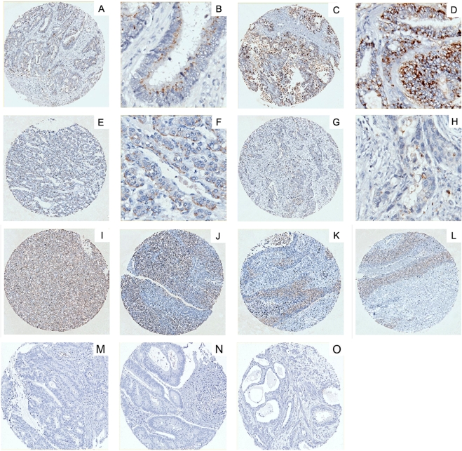
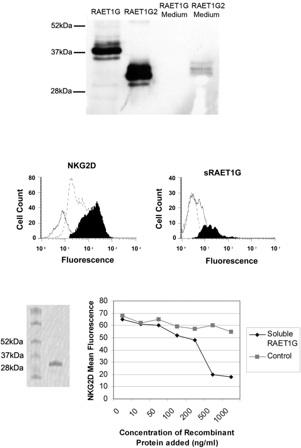
Methodology/principal findings: We pursue a number of approaches to characterise the expression, trafficking, and function of the two isoforms of ULBP5/RAET1G. We show that both transcripts are frequently expressed in cell lines derived from epithelial cancers, and in primary breast cancers. The full-length transcript, RAET1G1, is predicted to encode a molecule with transmembrane and cytoplasmic domains that are unique amongst NKG2D ligands. Using specific anti-RAET1G1 antiserum to stain tissue microarrays we show that RAET1G1 expression is highly restricted in normal tissues. RAET1G1 was expressed at a low level in normal gastrointestinal epithelial cells in a similar pattern to MICA. Both RAET1G1 and MICA showed increased expression in the gut of patients with celiac disease. In contrast to healthy tissues the RAET1G1 antiserum stained a wide variety or different primary tumour sections. Both endogenously expressed and transfected RAET1G1 was mainly found inside the cell, with a minority of the protein reaching the cell surface. Conversely the truncated splicing variant of RAET1G2 was shown to encode a soluble molecule that could be secreted from cells. Secreted RAET1G2 was shown to downregulate NKG2D receptor expression on NK cells and hence may represent a novel tumour immune evasion strategy.

Conclusions/significance: We demonstrate that the expression patterns of ULBP5RAET1G are very similar to the well-characterised NKG2D ligand, MICA. However the two isoforms of ULBP5/RAET1G have very different cellular localisations that are likely to reflect unique functionality.


Conflict of interest statement

Competing Interests: RAE and JT hold a patent on RAET1G.

Figures

Figure 1
Figure 1. Expression of two different isoforms of ULBP5/RAET1G.
(A) Two splicing variants of RAET1G have previously been identified. The full-length transcript, RAET1G1, is predicted to encode a protein with two MHC class I-related alpha domains, a transmembrane domain, and a cytoplasmic domain of 100 amino acids. RAET1G2 is predicted to encode a truncated protein that lacks the transmembrane and cytoplasmic domain. (B) Twenty epithelial cancer derived cell lines were analysed by RT-PCR for expression of full-length RAET1G1 transcript and the truncated splice variant RAET1G2. Both transcripts were present in most cell lines tested, at variable levels. (C) Expression of both variants was also very frequent in cDNA derived primary breast tumours. Expression of RAET1G1 and RAET1G2 appeared independent of p53 mutation status in a sample set of 12 tumours encoding wild-type p53 and 8 encoding mutated p53. Primers for GAPDH were used as a control for cDNA quantity.
Figure 2
Figure 2. Generation of specific anti-RAET1G1 antiserum.
(A) A rabbit polyclonal antiserum was raised against the cytoplasmic domain of RAET1G. By western blot a protein of approximately 70 kDa was recognised in Cos7 cells transfected with a construct encoding an RAET1G-GFP fusion protein, but not in untransfected Cos7 cells. (B) By confocal microscopy the anti-RAET1G antiserum specifically stains (red) Cos7 cells transfected with RAET1G-GFP (green), but not the highly related molecule ULBP2-GFP.
Figure 3
Figure 3. Immunohistochemical localisation of RAET1G1 in normal human tissues.
The anti-RAET1G antiserum was used to stain a tissue array of 344 cores from 172 donors that represented 35 normal tissues. Staining was very infrequently observed, but was present in (A) epithelial cells of colon (B) a minority of tubular cells in kidney, (C) majority of endocrine cells of the anterior pituitary and (D) cells lining thyroid follicles. The pituitary staining may represent true RAET1G1 expression or epitope cross reactivity, as has been shown to occur with other antibodies , . All other normal tissues were negative. Pre-immune serum did not stain any of these tissues; (E) colon, (F) kidney, (G) pituitary, and (H) thyroid.
Figure 4
Figure 4. Highly similar expression of RAET1G1 and MICA in healthy gut epithelium, and in patients with celiac disease.
(A) Expression of RAET1G and MICA in normal epithelial cells of the intestine. MICA was staining was with the monoclonal antibody SR99. Both proteins exhibited highly similar staining patterns of granular intracytoplasmic staining in epithelial cells of the brush border but not in the crypts. The MICA staining is essentially identical to previously published data where this pattern was indicative of low cell surface expression. (B) RAET1G and MICA staining in sections from the same patient with celiac disease. Both RAET1G and MICA showed increased and redistributed expression in the epithelial cell layer, with these cells exhibiting a more diffuse staining pattern. In the case of MICA this staining pattern has been shown to indicate higher cell surface expression of the protein. (C) Higher magnification image of the same section. Both RAET1G and MICA show some staining of intraintestinal cells that may be plasma cells. For each antibody four healthy controls and eight celiac disease patients were stained and the data are representative of the pattern seen in all samples.
Figure 5
Figure 5. RAET1G1 protein is poorly expressed at the cell surface.
(A) Stable cell lines expressing N-terminal GFP fusion proteins of ULBP2, and RAET1G1, were created in HT1080 cells. The transfectants expressed equivalent levels of transgene, as evident by total GFP fluorescence (open histogram), plotted versus untransfected HT1080 cells (gray shaded histogram). Cell surface expression was assessed by staining with an anti-GFP monoclonal antibody followed by an Alexa-Fluor 633 nm (far red) secondary antibody. The RAET1G1 transfectant had only a modest level of staining over untransfected cells, whereas the ULBP2 transfectant stained at a high level. This indicates that a much smaller percentage of RAET1G1-GFP transgene is present at the cell surface when compared to the closely related molecule ULBP2. (B) This observation was confirmed by confocal microscopy. The ULBP2-GFP transfectant showed clearly defined cell surface fluorescence in all cells, whereas cell surface RAET1G1-GFP could not be observed. (C) Stable transfectants were radiolabelled and chased for 180 minutes. Radiolabelled RAET1G1 and ULBP2 were then immunoprecipitated with an anti-GFP antibody. Endo H digests reveal that a substantial proportion of ULBP2 has acquired Endo H resistance after 180 minutes, and hence has trafficked to the cell surface. In contrast RAET1G1 remains Endo H sensitive.
Figure 6
Figure 6. Subcellular localisation of RAET1G1.
(A) By confocal microscopy, little RAET1G1 co-localised with MHC Class I (W6/32 antibody) at the cell surface, suggesting that the majority of RAET1G protein is found inside the cell. RAET1G1 does co-localise with calreticulin, a protein expressed predominately in the endoplasmic reticulum (ER). Co-localisation was also seen with the golgi apparatus marker GM130, as highlighted in inset. (C) Antibody internalisation experiment to prove that some RAET1G1 protein does reach the cell surface where it can be internalised into the endocytic pathway. Live cells were incubated with an anti-myc tag antibody prior to fixing and staining. Red represents myc tag staining, purple is staining with an antibody to the early endosome marker EEA1, and green is RAET1G1. Only RAET1G1 positive cells showed staining with the myc antibody; RAET1G1 negative cells only stained with anti-EEA1. In a magnified image EEA1, myc and RAET1G1 co-localised in vesicles (white, marked by arrows) showing that RAET1G1 is being internalised into early endosomes. Also, EEA1 negative, RAET1G1 positive, myc positive vesicles could be seen (yellow), and probably represent other compartments in the endocytic pathway. These data indicate that the vast majority of RAET1G1 protein is present in the ER and early golgi apparatus, and hence does not acquire Endo H resistance. A small minority of RAET1G1 can reach the cell surface, where it can be internalised into the endocytic pathway.
Figure 7
Figure 7. Immunohistochemical localisation of RAET1G in epithelial tumours.
A tissue microarray representing 35 different types of primary cancers was probed with the anti-RAET1G antiserum. In contrast normal tissue array widespread staining was observed, for example: (A) and (B) adenocarcinoma of colon, (C) and (D) adenocarcinoma of rectum, (E) and (F) follicular carcinoma of thyroid, (G) and (H) adenocarcinoma of stomach, (I) hepatoma, (J) serous carcinoma of ovary, (K) squamous cell carcinoma of lung and (L) squamous cell carcinoma of skin. All samples of these tumours showed staining, at varying intensity and number of positive cells. Again pre-immune serum did not show staining of tumour sections. (M), (N), and (O) are examples of negative staining in colonic tumours.
Figure 8
Figure 8. The truncated isoform, RAET1G2, can be secreted by cells and can downregulate NKG2D expression on NK cells.
(A) RAET1G2 transcript encodes a protein that is secreted from the cell. C-terminally V5 tagged RAET1G1 and RAET1G2 were transfected into Cos7 cells, RAET1G2 protein was readily detected in the tissue culture media of transfected cells by western blot with an anti-V5 antibody, whereas RAET1G1 was not demonstrated. (B) Soluble RAET1G2 down regulates NKG2D expression on natural killer lymphocytes. Isolated peripheral blood CD3− CD56+ NK cells were incubated for 24 hours at 37°C with culture supernatant from RAET1G1 transfected COS cells (solid black profile) or RAET1G2 transfected COS cells (open dashed profile) and examined for NKG2D expression levels using a PE-labelled anti-NKG2D antibody and flow cytometry. Supernatant from the RAET1G2 transfectant caused a substantial downregulation of NKG2D expression (mean fluorescence intensity = 41.75) compared with supernatant from the RAET1G1 transfectant (mean fluorescence intensity = 144.7). The solid white profile shows background staining with a PE-labelled mouse isotype control (mean fluorescence intensity = 8.1). (C) Soluble RAET1G2 binding to NK cells occurs rapidly but is lost within 24 hours of culture. V5 epitope tagged RAET1G2 was incubated at 37°C with isolated peripheral blood natural killer cells and assayed for binding after 1 hour (solid black profile) and 24 hours (open dashed profile) of incubation. RAET1G2 binding was detected by staining with anti-V5 antibody and PE-labelled goat anti-mouse second stage antibody followed by flow cytometric analysis. The solid white profile depicts background staining with the second stage antibody alone. (D) Coomassie stained SDS-PAGE gel of soluble recombinant RAET1G (rRAET1G). (E) rRAET1G down regulates NKG2D expression. Isolated peripheral blood CD3− CD56+ NK cells were incubated for 24 hours at 37°C with a range of concentrations of rRAET1G. Downregulation of NKG2D was observed at concentrations of rRAET1G as low as 100 ng/ml, but not in the presence of an irrelevant control his-tagged protein.

References

    1. Raulet DH. Roles of the NKG2D immunoreceptor and its ligands. Nat Rev Immunol. 2003;3:781–790. - PubMed
    1. Groh V, Bruhl A, El-Gabalawy H, Nelson JL, Spies T. Stimulation of T cell autoreactivity by anomalous expression of NKG2D and its MIC ligands in rheumatoid arthritis. Proc Natl Acad Sci USA. 2003;100:9452–9457. - PMC - PubMed
    1. Groh V, Smythe K, Dai Z, Spies T. Fas-ligand-mediated paracrine T cell regulation by the receptor NKG2D in tumor immunity. Nat Immunol. 2006;7:755–762. - PubMed
    1. Saez-Borderias A, Guma M, Angulo A, Bellosillo B, Pende D, et al. Expression and function of NKG2D in CD4+ T cells specific for human cytomegalovirus. Eur J Immunol. 2006;36:3198–3206. - PubMed
    1. Gonzalez S, Groh V, Spies T. Immunobiology of human NKG2D and its ligands. Curr Top Microbiol Immunol. 2006;298:121–138. - PubMed

Publication types

LinkOut - more resources