. 2009 Apr 17;284(16):10480-90.
doi: 10.1074/jbc.M809259200. Epub 2009 Feb 17.

Laminin receptor involvement in the anti-angiogenic activity of pigment epithelium-derived factor

Affiliations

Laminin receptor involvement in the anti-angiogenic activity of pigment epithelium-derived factor

Adrien Bernard et al. J Biol Chem. .

Abstract

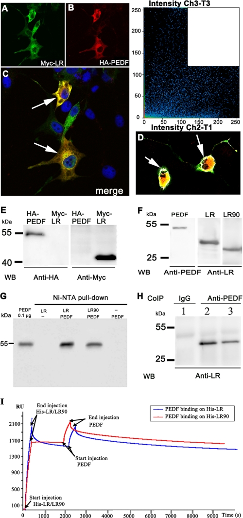
Pigment epithelium-derived factor (PEDF) is a multifunctional protein with neurotrophic, anti-oxidative, and anti-inflammatory properties. It is also one of the most potent endogenous inhibitors of angiogenesis, playing an important role in restricting tumor growth, invasion, and metastasis. Studies show that PEDF binds to cell surface proteins, but little is known about how it exerts its effects. Recently, research identified phospholipase A(2)/nutrin/patatin-like phospholipase domain-containing 2 as one PEDF receptor. To identify other receptors, we performed yeast two-hybrid screening using PEDF as bait and discovered that the non-integrin 37/67-kDa laminin receptor (LR) is another PEDF receptor. Co-immunoprecipitation, His tag pulldown, and surface plasmon resonance assays confirmed the interaction between PEDF and LR. Using the yeast two-hybrid method, we further restricted the LR-interacting domain on PEDF to a 34-amino acid (aa) peptide (aa 44-77) and the PEDF-interacting domain on LR to a 91-aa fragment (aa 120-210). A 25-mer peptide named P46 (aa 46-70), derived from 34-mer, interacts with LR in surface plasmon resonance assays and binds to endothelial cell (EC) membranes. This peptide induces EC apoptosis and inhibits EC migration, tube-like network formation in vitro, and retinal angiogenesis ex vivo, like PEDF. Our results suggest that LR is a real PEDF receptor that mediates PEDF angiogenesis inhibition.


Figures

FIGURE 1.
FIGURE 1.
Finding PEDF-LR interaction domains by yeast two-hybrid assay. A, identifying a direct LR-interacting domain on PEDF. Using full-length PEDF (aa 2–418) as bait, we identified the extracellular domain of LR (LRec 96–295) as a PEDF partner. We designated their interaction level as ++ and compared other experiments to this, labeling them as ++, +, and – depending on the speed of yeast growth and the blue staining of galactosidase activity. The corresponding amino acid numbers in each construct are indicated to the left of the figure. The interaction level is marked to the right of the figure. We identified one LR-interacting domain (aa 44–77) in N terminus. This fragment (aa 44–77) has also been identified as the anti-angiogenic peptide 34-mer (25). B, identifying the PEDF-interacting domain on LR. We tested the interactions between different fragment lengths of 67LR and full-length PEDF (2–418) or the 34-mer fragment. The PEDF-interacting domain on LR was localized to the region 120–210. The laminin/prion-binding domain (laminin/PrP BD) or peptide G (aa 157–180) alone was not sufficient for PEDF-LR interaction. However, deleting this region reduces PEDF-LR interaction. TM, transmembrane; HSPG BD, heparan sulfate proteoglycan-binding domain.
FIGURE 2.
FIGURE 2.
Co-localization of HA-PEDF and Myc-LR in cell cultures and PEDF-LR interaction in vitro. A and B, we immunostained Myc-LR and HA-PEDF co-transfected COS7 cells with anti-Myc antibody (green) and anti-HA antibody (red) 48 h after transfection. C, the merged image of A and B indicates the co-localization of PEDF and LR in the co-transfected cells (arrows). D, confocal microscopy image analysis of HA-PEDF and Myc-LR in COS7 cells with the software Coloc. The white color in the plot in the upper right corner corresponds to the co-localization of high density red color (Ch3-T3) and green color (Ch2-T1) (between 120 and 250) staining. This co-localization occurred mainly around the plasma membrane (arrows). E, Western blot (WB) analysis of COS7 cells transfected with anti-HA and anti-Myc antibodies. We found HA-PEDF at 55 kDa and Myc-LR at 40 kDa. The plasmids used in transfection are indicated at the top of the picture, and molecular mass markers are to the left. F, we confirmed PEDF expression in insect Sf9 cells and LR or LR90 in E. coli by Western blot, using anti-PEDF antibody and anti-LR antibody. G, PEDF was pulled down with Ni-NTA resin when His-tag LR or His-tag LR90 was present in the mixture. H, co-immunoprecipitation of PEDF and LR. Anti-LR only detected 40 kDa LR when we used anti-PEDF antibody (lanes 2 and 3) in the mixture containing full-length LR and PEDF, but not when we used control IgG (lane 1.). I, real time binding by SPR analyses of PEDF and LR interactions. We recorded sensograms with His-LR or His-LR90 immobilized on a NTA sensor Chip and PEDF using a BIAcore 3000 instrument and BIAevaluation software. We observed similar interactions (370 RU) between PEDF and LR or LR90.
FIGURE 3.
FIGURE 3.
Characterizing the 25-mer peptide that specifically interacts with LR. A, partial sequence (aa 1–135) of human PEDF. Secondary structure is indicated by H for helix and A for beta sheet. The 25-mer region (aa 46–70), with a helix-loop-helix is shown in red. This 25-mer is part of a previously described 34-mer by Filleur et al. (25). B, secondary structure of this 25-mer. C, in silico analysis with VMD software shows the potential location of 25-mer P46 within the three-dimensional structure of PEDF. D, SPR assays. His-LR was immobilized on a sensor Chip NTA to reach a response of between 1600 and 2000 RU. We analyzed the interaction between 25-mer and LR using a BIAcore 3000 instrument (BIAcore), according to the manufacturer's instructions. The results indicated a specific interaction between LR and 25-mer. We used peptide KAP3.1 as a negative control.
FIGURE 4.
FIGURE 4.
25-mer peptide F46 binds HuBMECs. A, Fluorescent-P46 (F46) binding to HuBMECs. We incubated HuBMECs with 200 nm F46 for 60 min at 4 °C. We used DAPI (blue) to label nuclei. B, rhodamin-labeled control peptide KAP3.1 (200 nm) did not bind to HuBMECs under the same condition as A. C, F46 binding to HuBMECs was inhibited when cells were preincubated with polyclonal 67LR antibody (ab711; Abcam). D, F46 binding to HuBMECs was decreased when cells were preincubated with monoclonal 67LR antibody (H-141; Santa Cruz). E, co-localization (white) of F46 and LR on the surface of the HuBMECs. We incubated F46 with cells, as described for A, and then incubated the cells with anti-67LR antibody (H-141; Santa Cruz) to label LR. White (arrow) demonstrates the co-localization of intense green staining (F46) and red staining (67LR). F, F46 binds specifically to HuBMEC membranes. We performed experiments as detailed for E, except for adding excess nonfluorescent P46 to the cells in the binding buffer. F46 staining (green) was barely detectable. Bar, 20 μm.
FIGURE 5.
FIGURE 5.
25-mer peptide P46 inhibits bFGF-induced angiogenesis on Matrigel. We seeded HuBMECs into 24-well plates coated with Matrigel and examined tube-like structure formation by phase contrast microscopy 24 h later. A, control: no factors in medium. B, we observed tube-like structure formation after 24 h of incubation with bFGF (20 ng/ml). C, PEDF (40 ng/ml) inhibits bFGF-induced tube-like structure formation. D, peptide P46 (200 nm) inhibits tube-like structure formation, just like PEDF. E, control peptide KAP3.1 (200 nm) does not inhibit tube-like structure formation. F, graphic representation of bFGF-induced tube-like structure formation in the presence of PEDF, P46, and KAP3.1, respectively. G, PEDF and P46 inhibit bFGF-induced angiogenesis in corneas. We show representative corneas here, indicating the number of positive corneas/total in parentheses.
FIGURE 6.
FIGURE 6.
PEDF and P46 peptide decrease cell motility in wound healing assays and promote apoptosis in endothelial cells. A, representative pictures of wound healing experiments. PEDF or the peptides used are shown at the top of the picture, and time is marked on the left. B, cell migration in the presence of PEDF, P46, and KAP3.1, respectively, at 4 and 8 h. PEDF and P46 decrease cell migration. C, apoptotic cells in different conditions. We starved HuBMECs in factor-free 0.2% FBS MEB2 overnight and incubated them in a starved medium with or without bFGF/VEGF growth factors (20 ng/ml each) and PEDF (40 ng/ml), P46 (200 nm), P326 (200 nm), and KAP3.1 (200 nm) for 24 h. bFGF/VEGF protects cells from apoptosis under starvation conditions. We counted the percentage of TUNEL-labeled cells by fluorescence microscopy, examining four random views/sample. We repeated the experiment three times.
FIGURE 7.
FIGURE 7.
PEDF and peptides promote apoptosis by regulating caspase-3 activity. A, PEDF and P46 increase caspase-3 activation. We transfected HuBMECs with either LR siRNA or scrambled siRNA. After 48 h, we treated cells with PEDF (40 ng/ml) or P46 (200 nm) for 30 min, 1 h, 3 h, or 6 h. We analyzed lysates by immunoblotting, using anti-LR, anti-active caspase-3, and anti-glyceraldehyde-3-phosphate dehydrogenase antibodies. We have shown a representative Western blot. B, reduced LR protein expression in LR siRNA-treated HuBMECs. We measured the relative 67LR expression and found a 60% decrease in LR protein in the LR siRNA-treated cells compared with the scrambled siRNA-treated cells. C and D, relative active caspase-3 levels. Graphs show the relative active caspase-3 level (compared with control cells) in the cells treated with LR or scrambled siRNA after 30 min, 1 h, 3 h, and 6 h of incubation with PEDF (C) or P46 (D) at 37 °C. Note the reduced active caspase-3 production in the siRNA-treated cells compared with the control cells in the presence of PEDF or P46. We repeated the experiment once.

References

    1. Tombran-Tink, J., Chader, G. G., and Johnson, L. V. (1991) Exp. Eye Res. 53 411–414 - PubMed
    1. Tombran-Tink, J., Mazuruk, K., Rodriguez, I. R., Chung, D., Linker, T., Englander, E., and Chader, G. J. (1996) Mol. Vis. 2 11. - PubMed
    1. Bouck, N. (2002) Trends Mol. Med. 8 330–334 - PubMed
    1. Tombran-Tink, J., and Barnstable, C. J. (2003) Nat. Rev. Neurosci. 4 628–636 - PubMed
    1. Becerra, S. P. (2006) Exp. Eye Res. 82 739–740 - PubMed

Publication types

MeSH terms