. 2009 Apr 24;284(17):11706-16.
doi: 10.1074/jbc.M805990200. Epub 2009 Feb 17.

Synphilin-1A inhibits seven in absentia homolog (SIAH) and modulates alpha-synuclein monoubiquitylation and inclusion formation

Affiliations

Synphilin-1A inhibits seven in absentia homolog (SIAH) and modulates alpha-synuclein monoubiquitylation and inclusion formation

Raymonde Szargel et al. J Biol Chem. .

Abstract

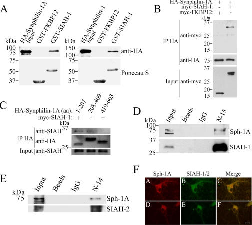
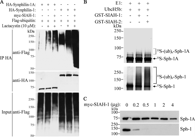
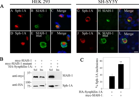
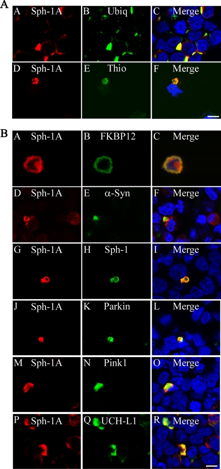
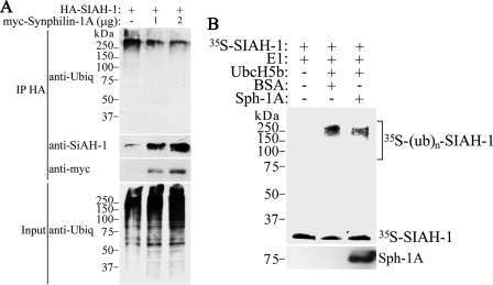
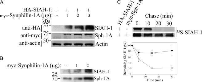
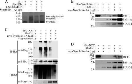
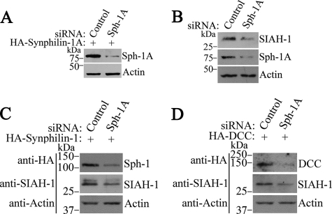
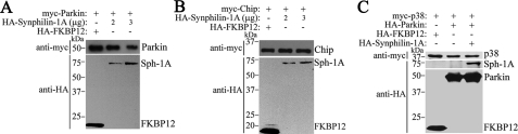
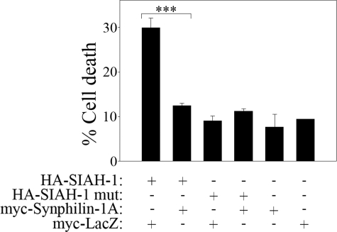
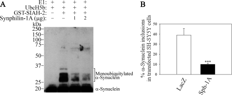
Parkinson disease (PD) is characterized by the presence of ubiquitylated inclusions and the death of dopaminergic neurons. Seven in absentia homolog (SIAH) is a ubiquitin-ligase that ubiquitylates alpha-synuclein and synphilin-1 and is present in Lewy bodies of PD patients. Understanding the mechanisms that regulate the ubiquitylation of PD-related proteins might shed light on the events involved in the formation of Lewy bodies and death of neurons. We show in this study that the recently described synphilin-1 isoform, synphilin-1A, interacts in vitro and in vivo with the ubiquitin-protein isopeptide ligase SIAH and regulates its activity toward alpha-synuclein and synphilin-1. SIAH promotes limited ubiquitylation of synphilin-1A that does not lead to its degradation by the proteasome. SIAH also increases the formation of synphilin-1A inclusions in the presence of proteasome inhibitors, supporting the participation of ubiquitylated synphilin-1A in the formation of Lewy body-like inclusions. Synphilin-1A/SIAH inclusions recruit PD-related proteins, such as alpha-synuclein, synphilin-1, Parkin, PINK1, and UCH-L1. We found that synphilin-1A robustly increases the steady-state levels of SIAH by decreasing its auto-ubiquitylation and degradation. In addition, synphilin-1A blocks the ubiquitylation and degradation of the SIAH substrates synphilin-1 and deleted in colon cancer protein. Furthermore, synphilin-1A strongly decreases the monoubiquitylation of alpha-synuclein by SIAH and the formation of alpha-synuclein inclusions, supporting a role for monoubiquitylation in alpha-synuclein inclusion formation. Our results suggest a novel function for synphilin-1A as a regulator of SIAH activity and formation of Lewy body-like inclusions.


Figures

FIGURE 1.
FIGURE 1.
SIAH interacts with synphilin-1A in vitro and in vivo. A, left panel, extract of HEK293 cells transfected with HA-synphilin-1A was incubated with the indicated GST fusion proteins. Binding was analyzed by using an anti-HA antibody. Right panel, extract of HEK293 cells transfected with HA-synphilin-1 was incubated with the indicated GST fusion proteins. Binding was analyzed using anti-HA antibody. The total amount of GST fusion proteins used in the experiments was determined by staining the membranes with Ponceau S (lower panels). B, SIAH-1 co-immunoprecipitates with synphilin-1A from co-transfected HEK293 cells. HA-synphilin-1A was immunoprecipitated (IP) from extracts of HEK293 cells using an anti-HA antibody. Co-immunoprecipitation was determined by Western blot using an anti-Myc antibody. C, mapping the SIAH binding region in synphilin-1A. HA-synphilin-1A fragments were immunoprecipitated from extracts of co-transfected HEK293 cells using an anti-HA antibody. The co-immunoprecipitation of SIAH-1 with synphilin-1A fragments was determined by using the anti-SIAH antibody 990. The detection of immunoprecipitated synphilin-1A fragments was done using an anti-HA antibody. aa, amino acids. D, synphilin-1A (Sph-1A) co-immunoprecipitates with endogenous SIAH-1. SIAH-1 was immunoprecipitated from rat brain homogenate using the anti-SIAH-1 antibody (N-15), and detection of co-immunoprecipitation was carried out using anti-synphilin-1A antibody (25). The detection of immunoprecipitated-SIAH-1 was done using the anti-SIAH-1 antibody (990) (8). E, synphilin-1A also co-immunoprecipitates with endogenous SIAH-2. SIAH-2 was immunoprecipitated from rat brain homogenate using the anti-SIAH-2 antibody (N-14), and detection of co-immunoprecipitation was carried out using anti-synphilin-1A antibody. The detection of immunoprecipitated-SIAH-2 was done using the anti-SIAH-1/2 antibody (H-18). F, rat cortical neurons grown for 14 days in vitro were immunolabeled against synphilin-1A (panels A and D), SIAH-1 with N-15 antibody (panel B), and SIAH-1/2 with H-18 antibody (panel E). The co-localization of endogenous synphilin-1A and SIAH in the cytoplasm of neurons, including cell bodies and neuronal processes, is observed in the merge pictures (panels C and F). Scale bar, 20 μm.
FIGURE 2.
FIGURE 2.
SIAH promotes slight ubiquitylation and degradation of synphilin-1A. A, HEK293 cells were co-transfected with HA-synphilin-1A, FLAG-ubiquitin, in the absence or in the presence of myc-SIAH-1. As control, co-transfections were carried out with synphilin-1 instead of synphilin-1A. Cells were incubated 12 h with 10 μm lactacystin, and HA-synphilin-1A and HA-synphilin-1 were immunoprecipitated (IP) with anti-HA antibody. Ubiquitylation of synphilin-1A and synphilin-1 was detected by Western blot using anti-FLAG antibody. The middle panel shows the levels of immunoprecipitated synphilin-1A and synphilin-1 using anti-HA antibody. The lower panel shows the total ubiquitylation levels of co-transfected cells with anti-FLAG antibody. B, in vitro translated 35S-synphilin-1A was incubated with recombinant SIAH-1 or SIAH-2, UbcH5b, ubiquitin, and the other purified components of the ubiquitin system. The levels of 35S-synphilin-1A in vitro ubiquitylation were determined by PhosphorImager analysis (upper panel). As control, in vitro translated 35S-synphilin-1 was incubated with recombinant SIAH-1 or SIAH-2 under the same conditions as for 35S-synphilin-1A, and the levels of 35S-synphilin-1 ubiquitylation were determined by PhosphorImager analysis (lower panel). E1, ubiquitin-activating enzyme. C, effect of SIAH-1 on synphilin-1A (Sph-1A) steady-state levels. HEK293 cells were transfected with HA-synphilin-1A and myc-SIAH-1. HA-synphilin-1A and HA-synphilin-1 from total cell lysates were detected by Western blot using anti-HA antibody.
FIGURE 3.
FIGURE 3.
SIAH is recruited to synphilin-1A inclusions. A, immunofluorescence of transfected HEK293 (left panels) and SH-SY5Y (right panels) cells reveals the co-localization of myc-SIAH-1 but not myc-SIAH-1 catalytically inactive mutant (C55A,C59H,C72S) with HA-synphilin-1A inclusions. Immunocytochemistry was carried out with anti-HA (red) and anti-Myc antibody (green). Nuclei were revealed with TOPRO-3. Scale bar, 25 μm. B, synphilin-1A (Sph-1A) recruits wild-type SIAH-1 but not the catalytically inactive mutant to the Triton X-100-insoluble fraction. HEK293 cells transfected with HA-synphilin-1A and myc-SIAH-1 or myc-SIAH-1 inactive mutant were lysed, and cell lysates were divided into Triton-soluble (Sol) and Triton-insoluble (Ins) fractions. The distribution of SIAH-1 (wild-type and inactive mutant) between soluble and insoluble fractions was determined using anti-Myc antibody (upper panels). The distribution of synphilin-1A between the same fractions was determined using anti-HA antibody. C, quantification of synphilin-1A inclusion formation in SH-SY5Y cells transfected with HA-synphilin-1A, in the absence or in the presence of myc-SIAH-1. Cells were incubated 12 h with 10 μm lactacystin prior to the immunocytochemistry assay. Error bars represent standard error of five independent experiments. **, significantly different from control at p < 0.01.
FIGURE 4.
FIGURE 4.
Co-localization of PD-related proteins with synphilin-1A/SIAH inclusions. A, SH-SY5Y cells were co-transfected with HA-synphilin-1A (Sph-1A), untagged SIAH-1, and FLAG-ubiquitin (Ubiq). Immunocytochemistry was carried out with anti-HA (red) and anti-FLAG or thioflavine (Thio) S (14) (green). Nuclei were stained with TOPRO-3. Scale bar, 25 μm. B, SH-SY5Y cells were co-transfected with HA-synphilin-1A, untagged SIAH-1, and myc-FKBP12, myc-α-synuclein (α-Syn), myc-synphilin-1, myc-Parkin, myc-PINK1, or myc-UCH-L1. Immunocytochemistry was carried out with anti-HA (red) and anti-Myc (green). Nuclei were stained with TOPRO-3. Scale bar, 25 μm. The figures are representative of 3-6 independent experiments.
FIGURE 5.
FIGURE 5.
Synphilin-1A decreases the ubiquitylation of SIAH-1. A, HEK293 cells were transfected with HA-SIAH-1 and increasing amounts of myc-synphilin-1A. Cells were incubated 12 h with 10 μm lactacystin. HA-SIAH-1 was immunoprecipitated (IP) with anti-HA antibody, and ubiquitylated SIAH-1 was detected by Western blot using anti-ubiquitin (Ubiq) antibody. The amount of immunoprecipitated SIAH-1 was determined using anti-SIAH-1 antibody N-15 (2nd panel). The levels of synphilin-1A (Sph-1A) were determined with anti-Myc antibody (3rd panel). B, in vitro translated SIAH-1 was incubated with UbcH5b, ubiquitin, and the other purified components of the ubiquitin system in the absence or in the presence of recombinant synphilin-1A. The levels of SIAH-1 ubiquitylation were determined by PhosphorImager analysis. The amount of synphilin-1A added was determined by Western blot using anti-synphilin-1A antibody (lower panel). BSA, bovine serum albumin.
FIGURE 6.
FIGURE 6.
Synphilin-1A inhibits the degradation of SIAH-1. A, HEK293 cells were co-transfected with HA-SIAH-1 and increasing amounts of myc-synphilin-1A. HA-SIAH-1 steady-state levels from total cell lysates were detected by Western blot with anti-HA antibody. The expression levels of synphilin-1A were determined using anti-Myc antibody. Loading control was monitored with anti-actin antibody (lower panel). B, HEK293 cells were transfected with increasing amounts of myc-synphilin-1A. Steady-state levels of endogenous SIAH-1 from total cell lysates were determined using anti-SIAH-1 antibody (N-15). The expression levels of synphilin-1A were determined using anti-Myc antibody. C, synphilin-1A (Sph-1A) increases the half-life of SIAH-1. Transfected HEK293 cells were chased for the indicated time points, and HA-SIAH-1 was immunoprecipitated using anti-HA antibody. Immunoprecipitates were analyzed by SDS-PAGE and autoradiography (upper panel). Graph shows the quantification of remaining SIAH-1 at indicated time points. Error bars represent standard error of three independent experiments (lower panel).
FIGURE 7.
FIGURE 7.
Synphilin-1A decreases the ubiquitylation and degradation of the SIAH-1 substrates synphilin-1 and DCC. A, in vitro translated synphilin-1 was incubated with increasing amounts of synphilin-1A, UbcH5b, ubiquitin, and the other purified components of the ubiquitin system, in the absence or in the presence of SIAH-1. The levels of synphilin-1 ubiquitylation were determined by PhosphorImager analysis. E1, ubiquitin-activating enzyme. B, HEK293 cells were transfected with HA-synphilin-1, untagged SIAH-1, and increasing amounts of synphilin-1A. HA-synphilin-1 from total cell lysates was detected by Western blot using anti-HA antibody (upper panel). The expression levels of synphilin-1A (Sph-1A) and SIAH-1 were determined with anti-Myc and anti-SIAH-1 (N-15) antibodies, respectively. C, HEK293 cells were transfected with HA-DCC and increasing amounts of myc-synphilin-1A. Cells were incubated 12 h with 10 μm lactacystin. HA-DCC was immunoprecipitated with anti-HA antibody, and ubiquitylated DCC was detected by Western blot using anti-FLAG antibody. The amount of immunoprecipitated DCC was determined using anti-HA antibody (2nd panel). The levels of synphilin-1A were determined with anti-Myc antibody (4th panel). D, HEK293 cells were transfected with HA-DCC, untagged SIAH-1, and increasing amounts of synphilin-1A. HA-DCC from total cell lysates was detected by Western blot using anti-HA antibody (upper panel). The expression levels of synphilin-1A and SIAH-1 were determined with anti-Myc and anti-SIAH-1 (N-15) antibodies, respectively.
FIGURE 8.
FIGURE 8.
Reduction of synphilin-1A expression decreases the steady-state levels of SIAH-1 and its substrates synphilin-1 and DCC. A, SH-SY5Y cells were transfected with HA-synphilin-1A, in the presence of siRNA control or siRNA to synphilin-1A (Sph-1A). HA-synphilin-1A from total cell lysates was detected by Western blot using anti-HA antibody (upper panel). B, SH-SY5Y cells were transfected with siRNA control or siRNA to synphilin-1A. The endogenous levels of SIAH-1 from total cell lysates were detected by Western blot using the 990 anti-SIAH-1 antibody (8) (upper panel). Middle panel shows the levels of endogenous synphilin-1A with anti-synphilin-1A antibody. C, SH-SY5Y cells were transfected with HA-synphilin-1, in the presence of siRNA control or siRNA to synphilin-1A. HA-synphilin-1 from total cell lysates was detected by Western blot using anti-HA antibody (upper panel). Middle panel shows the levels of endogenous SIAH-1 with 990 anti-SIAH-1 antibody. D, SH-SY5Y cells were transfected with HA-DCC, in the presence of siRNA control or siRNA to synphilin-1A. HA-DCC from total cell lysates was detected by Western blot using anti-HA antibody (upper panel). Middle panel shows the levels of endogenous SIAH-1 with 990 anti-SIAH-1 antibody. Lower panels in A-D show the loading control of protein extracts determined by incubating the membranes with anti-actin antibody.
FIGURE 9.
FIGURE 9.
Synphilin-1A does not interfere with the steady-state levels of Parkin and its substrate p38 as well as with CHIP. A, HEK293 cells were transfected with myc-Parkin and increasing amounts of synphilin-1A (Sph-1A) or HA-FKBP12. myc-Parkin from total cell lysates was detected by Western blot using anti-Myc antibody (upper panel). The expression levels of synphilin-1A and FKBP12 were determined with anti-HA. B, HEK293 cells were transfected with myc-CHIP and increasing amounts of synphilin-1A or HA-FKBP12. myc-CHIP from total cell lysates was detected by Western blot using anti-Myc antibody (upper panel). The expression levels of synphilin-1A and FKBP12 were determined with anti-HA. C, HEK293 cells were transfected with myc-p38, HA-Parkin, and HA-FKBP12 in the absence or in the presence of HA-synphilin-1A. myc-p38 from total cell lysates was detected by Western blot using anti-Myc antibody (upper panel). The expression levels of Parkin, synphilin-1A, and FKBP12 were determined with anti-HA.
FIGURE 10.
FIGURE 10.
Synphilin-1A decreases the toxicity of SIAH. HEK293 cells were transfected with HA-SIAH-1, HA-SIAH-1 C55A,H59A,C72S (ligase-inactive mutant (mut)) (8), myc-synphilin-1A, and myc-LacZ. Immunocytochemistry was carried out using anti-HA and anti-Myc antibodies, and cell death was determined by quantifying the nuclear fragmentation and condensation of transfected HEK293 cells with Hoechst 33342. Error bars represent standard error of 4-7 independent experiments. ***, significantly different from vehicle control at p < 0.001.
FIGURE 11.
FIGURE 11.
Synphilin-1A decreases the monoubiquitylation of α-synuclein promoted by SIAH and the formation of monoubiquitylated α-synuclein inclusions. A, His-α-synuclein was incubated with increasing amounts of synphilin-1A (Sph-1A), UbcH5b, and the other components of the ubiquitin system, in the absence or in the presence of recombinant SIAH-2. His-α-synuclein ubiquitylation was determined by Western blot using an antibody to α-synuclein. E1, ubiquitin-activating enzyme. B, SH-SY5Y cells were transfected with HA-α-synuclein, untagged SIAH-2, in the presence of myc-synphilin-1A or LacZ Myc. Cells were incubated for 12 h with a combination of 10 μm lactacystin, 10 mm NH4Cl, and 10 mm 3-MA. Immunocytochemistry was carried out using anti-HA and anti-Myc antibodies, and the percent of α-synuclein inclusions was quantified in transfected SH-SY5Y cells. Error bars represent standard error of five independent experiments. ***, significantly different from vehicle control at p < 0.001.

Similar articles

Cited by

References

    1. Moore, D. J., West, A. B., Dawson, V. L., and Dawson, T. M. (2005) Annu. Rev. Neurosci. 28, 57-87 - PubMed
    1. Hardy, J., Cai, H., Cookson, M. R., Gwinn-Hardy, K., and Singleton, A. (2006) Ann. Neurol. 60, 389-398 - PubMed
    1. Spillantini, M. G., Crowther, R. A., Jakes, R., Hasegawa, M., and Goedert, M. (1998) Proc. Natl. Acad. Sci. U. S. A. 95, 6469-6473 - PMC - PubMed
    1. Lee, V. M., and Trojanowski, J. Q. (2006) Neuron 52, 33-38 - PubMed
    1. McNaught, K. S., and Jenner, P. (2001) Neurosci. Lett. 297, 191-194 - PubMed

Publication types

MeSH terms

LinkOut - more resources