. 2009 May;251(2):429-38.
doi: 10.1148/radiol.2511080539.

Atherosclerosis and matrix metalloproteinases: experimental molecular MR imaging in vivo

Affiliations

Atherosclerosis and matrix metalloproteinases: experimental molecular MR imaging in vivo

Vardan Amirbekian et al. Radiology. 2009 May.

Abstract

Purpose: To evaluate the capability of P947, a magnetic resonance (MR) imaging contrast agent that molecularly targets matrix metalloproteinases (MMPs), to aid detection and imaging of MMPs in atherosclerotic lesions in vivo; its specificity compared with that of P1135; expression and distribution of MMPs in atherosclerotic vessels; and in vivo distribution and molecular localization of fluorescent europium (Eu) P947.

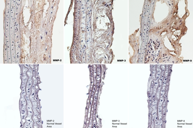
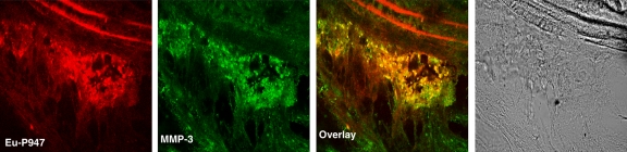
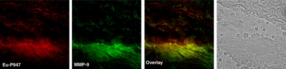
Materials and methods: The Animal Care and Use Committee approved all experiments. P947 was synthesized by attaching a gadolinium chelate (1,4,7,10-tetraazacyclododecane-N,N',N'',N'''-tetraacetic acid) to a peptide that specifically binds MMPs. Scrambled form of P947 (P1135) was synthesized by replacing the targeting moiety of P947 with a scrambled peptide lacking the ability to bind MMPs. P947, P1135, and gadoterate meglumine were injected into atherosclerotic apolipoprotein E-deficient and wild-type mice. The aortic MR imaging enhancement produced by the contrast agents was measured at different times and was compared by using one-way analysis of variance. MMP expression was investigated in the aortas by using MMP immunostaining and in situ MMP zymography. A fluorescent form of P947 (Eu-P947) was synthesized to compare the in vivo distribution of the contrast agent (Eu-P947) with specific MMP immunofluorescent staining.

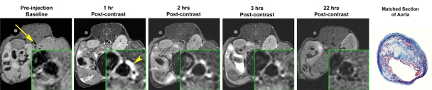
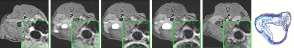
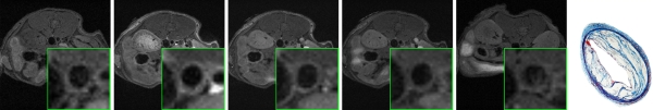
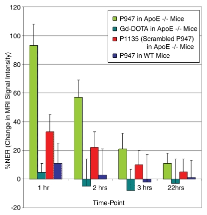
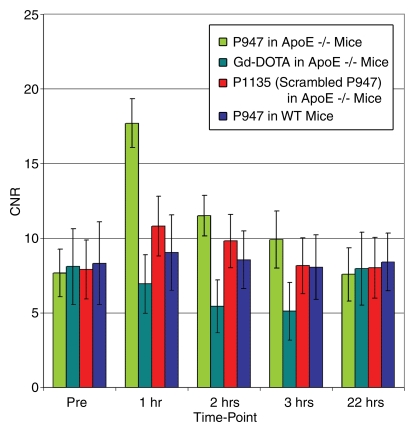
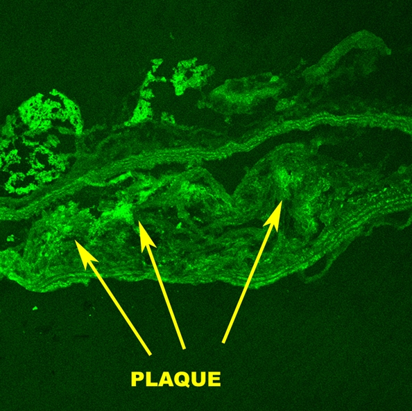
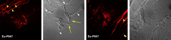
Results: MMP-targeted P947 facilitated a 93% increase (P < .001) in MR image signal intensity (contrast-to-noise ratio [CNR], 17.7 compared with 7.7; P < .001) of atherosclerotic lesions in vivo. Nontargeted P1135 (scrambled P947) provided 33% MR image enhancement (CNR, 10.8), whereas gadoterate meglumine provided 5% (CNR, 6.9). Confocal laser scanning microscopy demonstrated colocalization between fluorescent Eu-P947 and MMPs in atherosclerotic plaques. Eu-P947 was particularly present in the fibrous cap region of plaques.

Conclusion: P947 improved MR imaging for atherosclerosis through MMP-specific targeting. The results were validated and provide support for further assessment of P947 as a potential tool for the identification of unstable atherosclerosis.


Figures

Figure 1a:
Figure 1a:
In vivo MR images acquired in atherosclerotic ApoE-deficient mice. (a) At baseline (before injection) and up to 22 hours after injection of MMP-targeted P947. (b) With untargeted P1135 (scrambled form of P947). (c) With standard nontargeted gadoterate meglumine. Insets are enlargements of aorta (arrow). Arrowhead points to MR inset of atherosclerotic aorta after injection with P947. (a–c) Farthest right images show hematoxylin-eosin–stained sections of aorta at identical anatomic level as MR images from same animal (17).
Figure 1b:
Figure 1b:
In vivo MR images acquired in atherosclerotic ApoE-deficient mice. (a) At baseline (before injection) and up to 22 hours after injection of MMP-targeted P947. (b) With untargeted P1135 (scrambled form of P947). (c) With standard nontargeted gadoterate meglumine. Insets are enlargements of aorta (arrow). Arrowhead points to MR inset of atherosclerotic aorta after injection with P947. (a–c) Farthest right images show hematoxylin-eosin–stained sections of aorta at identical anatomic level as MR images from same animal (17).
Figure 1c:
Figure 1c:
In vivo MR images acquired in atherosclerotic ApoE-deficient mice. (a) At baseline (before injection) and up to 22 hours after injection of MMP-targeted P947. (b) With untargeted P1135 (scrambled form of P947). (c) With standard nontargeted gadoterate meglumine. Insets are enlargements of aorta (arrow). Arrowhead points to MR inset of atherosclerotic aorta after injection with P947. (a–c) Farthest right images show hematoxylin-eosin–stained sections of aorta at identical anatomic level as MR images from same animal (17).
Figure 2a:
Figure 2a:
Graphs depict quantitative temporal MR imaging results. In vivo MR image SI of aortic wall at different times measured with (a) percentage NER and (b) CNR in ApoE-deficient mice injected with MMP-targeted P947, gadoterate meglumine (Gd-DOTA), and untargeted P1135 and in WT mice injected with P947. In ApoE-deficient mice at 1 hour after injection, MMP-targeted P947 showed CNR of 17.7 (percentage NER, 93%); with gadoterate meglumine, CNR was 6.9 (percentage NER, 5%), with untargeted P1135, CNR was 10.8 (percentage NER, 33%), and in WT mice injected with P947, CNR was 8.3 (percentage NER, 11%).
Figure 2b:
Figure 2b:
Graphs depict quantitative temporal MR imaging results. In vivo MR image SI of aortic wall at different times measured with (a) percentage NER and (b) CNR in ApoE-deficient mice injected with MMP-targeted P947, gadoterate meglumine (Gd-DOTA), and untargeted P1135 and in WT mice injected with P947. In ApoE-deficient mice at 1 hour after injection, MMP-targeted P947 showed CNR of 17.7 (percentage NER, 93%); with gadoterate meglumine, CNR was 6.9 (percentage NER, 5%), with untargeted P1135, CNR was 10.8 (percentage NER, 33%), and in WT mice injected with P947, CNR was 8.3 (percentage NER, 11%).
Figure 3:
Figure 3:
Light microscopic images show specific MMP immunostaining of atherosclerotic aortic lesions from ApoE-deficient mice. Top: Increased expression of MMP-2, MMP-3, and MMP-9. Bottom: Normal vessel areas. Sections were taken from aortas of mice used in in vivo MR imaging experiments.
Figure 4a:
Figure 4a:
Fluorescence microscopic images from MMP in situ zymography of mouse aortas demonstrate increased MMP gelatinolytic activity in atherosclerotic sections. (a) Control section that was incubated with gel alone shows that there is negligible intrinsic fluorescence. (b) Atherosclerotic aortic section incubated with gel and dye that fluoresced only after cleavage by MMPs (arrows point to atherosclerotic plaque). (c) Atherosclerotic section that was incubated with gel, dye, and ethylenediaminetetraacetic acid, which blocks MMP activity, demonstrates that most of fluorescence seen on b was caused by active MMP cleavage of dye. All sections taken from mice used in in vivo MR imaging experiments.
Figure 4b:
Figure 4b:
Fluorescence microscopic images from MMP in situ zymography of mouse aortas demonstrate increased MMP gelatinolytic activity in atherosclerotic sections. (a) Control section that was incubated with gel alone shows that there is negligible intrinsic fluorescence. (b) Atherosclerotic aortic section incubated with gel and dye that fluoresced only after cleavage by MMPs (arrows point to atherosclerotic plaque). (c) Atherosclerotic section that was incubated with gel, dye, and ethylenediaminetetraacetic acid, which blocks MMP activity, demonstrates that most of fluorescence seen on b was caused by active MMP cleavage of dye. All sections taken from mice used in in vivo MR imaging experiments.
Figure 4c:
Figure 4c:
Fluorescence microscopic images from MMP in situ zymography of mouse aortas demonstrate increased MMP gelatinolytic activity in atherosclerotic sections. (a) Control section that was incubated with gel alone shows that there is negligible intrinsic fluorescence. (b) Atherosclerotic aortic section incubated with gel and dye that fluoresced only after cleavage by MMPs (arrows point to atherosclerotic plaque). (c) Atherosclerotic section that was incubated with gel, dye, and ethylenediaminetetraacetic acid, which blocks MMP activity, demonstrates that most of fluorescence seen on b was caused by active MMP cleavage of dye. All sections taken from mice used in in vivo MR imaging experiments.
Figure 5a:
Figure 5a:
Confocal laser scanning fluorescence microscopic images show Eu-P947 localization alone and colocalization with MMPs. Aortic sections from ApoE-deficient mice injected in vivo with Eu-P947 show localization of fluorescent Eu-P947 (red) in atherosclerotic plaques (far left and right images on a), alternating with their corresponding differential interference contrast light microscopic images (left and far right images on a). Yellow arrowheads indicate elastic lamina. Arrows point to fibrous cap in light microscopic image and show possible accumulation of P947 in fibrous cap (far left image on a). Fluorescent immunostaining for MMP-2, MMP-3, and MMP-9 (green on b–d, respectively) in aortic sections obtained from ApoE-deficient mice injected with fluorescent Eu-P947 (red on b–d). Overlaid images at right on b, c, and d show substantial overlap (yellow areas) between Eu-P947 and MMP-2, MMP-3, and MMP-9, respectively. On b and c, differential interference contrast light microscopic images at right are of respective sections at left. White arrowheads indicate general borders of atherosclerotic plaques on a and b.
Figure 5b:
Figure 5b:
Confocal laser scanning fluorescence microscopic images show Eu-P947 localization alone and colocalization with MMPs. Aortic sections from ApoE-deficient mice injected in vivo with Eu-P947 show localization of fluorescent Eu-P947 (red) in atherosclerotic plaques (far left and right images on a), alternating with their corresponding differential interference contrast light microscopic images (left and far right images on a). Yellow arrowheads indicate elastic lamina. Arrows point to fibrous cap in light microscopic image and show possible accumulation of P947 in fibrous cap (far left image on a). Fluorescent immunostaining for MMP-2, MMP-3, and MMP-9 (green on b–d, respectively) in aortic sections obtained from ApoE-deficient mice injected with fluorescent Eu-P947 (red on b–d). Overlaid images at right on b, c, and d show substantial overlap (yellow areas) between Eu-P947 and MMP-2, MMP-3, and MMP-9, respectively. On b and c, differential interference contrast light microscopic images at right are of respective sections at left. White arrowheads indicate general borders of atherosclerotic plaques on a and b.
Figure 5c:
Figure 5c:
Confocal laser scanning fluorescence microscopic images show Eu-P947 localization alone and colocalization with MMPs. Aortic sections from ApoE-deficient mice injected in vivo with Eu-P947 show localization of fluorescent Eu-P947 (red) in atherosclerotic plaques (far left and right images on a), alternating with their corresponding differential interference contrast light microscopic images (left and far right images on a). Yellow arrowheads indicate elastic lamina. Arrows point to fibrous cap in light microscopic image and show possible accumulation of P947 in fibrous cap (far left image on a). Fluorescent immunostaining for MMP-2, MMP-3, and MMP-9 (green on b–d, respectively) in aortic sections obtained from ApoE-deficient mice injected with fluorescent Eu-P947 (red on b–d). Overlaid images at right on b, c, and d show substantial overlap (yellow areas) between Eu-P947 and MMP-2, MMP-3, and MMP-9, respectively. On b and c, differential interference contrast light microscopic images at right are of respective sections at left. White arrowheads indicate general borders of atherosclerotic plaques on a and b.
Figure 5d:
Figure 5d:
Confocal laser scanning fluorescence microscopic images show Eu-P947 localization alone and colocalization with MMPs. Aortic sections from ApoE-deficient mice injected in vivo with Eu-P947 show localization of fluorescent Eu-P947 (red) in atherosclerotic plaques (far left and right images on a), alternating with their corresponding differential interference contrast light microscopic images (left and far right images on a). Yellow arrowheads indicate elastic lamina. Arrows point to fibrous cap in light microscopic image and show possible accumulation of P947 in fibrous cap (far left image on a). Fluorescent immunostaining for MMP-2, MMP-3, and MMP-9 (green on b–d, respectively) in aortic sections obtained from ApoE-deficient mice injected with fluorescent Eu-P947 (red on b–d). Overlaid images at right on b, c, and d show substantial overlap (yellow areas) between Eu-P947 and MMP-2, MMP-3, and MMP-9, respectively. On b and c, differential interference contrast light microscopic images at right are of respective sections at left. White arrowheads indicate general borders of atherosclerotic plaques on a and b.

Comment in

Similar articles

Cited by

References

    1. Fuster V, Moreno PR, Fayad ZA, Corti R, Badimon JJ. Atherothrombosis and high-risk plaque. I. Evolving concepts. J Am Coll Cardiol 2005;46:937–954. - PubMed
    1. Heart disease and stroke statistics—2006 update. American Heart Association Web site. http://www.americanheart.org/downloadable/heart/1136308648540Statupdate2.... Published 2006. Accessed September 23, 2006.
    1. Tunstall-Pedoe H. Preventing chronic diseases: a vital investment. WHO Global report. Geneva, Switzerland: World Health Organization, 2005. http://www.who.int/chp/chronic_disease_report/contents/part1.pdf. Accessed January 6, 2008.
    1. Anderson GF, Chu E. Expanding priorities: confronting chronic disease in countries with low income. N Engl J Med 2007;356:209–211. - PubMed
    1. Naghavi M, Libby P, Falk E, et al. From vulnerable plaque to vulnerable patient: a call for new definitions and risk assessment strategies. II. Circulation 2003;108:1772–1778. - PubMed

Publication types

MeSH terms