. 2009 Apr 17;284(16):10694-705.
doi: 10.1074/jbc.M807136200. Epub 2009 Feb 18.

Inositol 1,4,5-triphosphate receptor-binding protein released with inositol 1,4,5-triphosphate (IRBIT) associates with components of the mRNA 3' processing machinery in a phosphorylation-dependent manner and inhibits polyadenylation

Affiliations

Inositol 1,4,5-triphosphate receptor-binding protein released with inositol 1,4,5-triphosphate (IRBIT) associates with components of the mRNA 3' processing machinery in a phosphorylation-dependent manner and inhibits polyadenylation

Hélène Kiefer et al. J Biol Chem. .

Abstract

IRBIT is a recently identified protein that modulates the activities of both inositol 1,4,5-triphosphate receptor and pancreas-type Na(+)/HCO(3)(-) cotransporter 1, and the multisite phosphorylation of IRBIT is required for achieving this modulatory action. Here, we report the identification of the cleavage and polyadenylation specificity factor (CPSF), which is a multi-protein complex involved in 3' processing of mRNA precursors, as an additional binding partner for IRBIT. We found that IRBIT interacted with CPSF and was recruited to an exogenous polyadenylation signal-containing RNA. The main target for IRBIT in CPSF was Fip1 subunit, and the phosphorylation of the serine-rich region of IRBIT was required both for direct association with Fip1 in vitro and for redistribution of Fip1 into the cytoplasm of intact cells. Furthermore, tert-butylhydroquinone (tBHQ), an agent that induces oxidative stress, increased the phosphorylation level of IRBIT in vivo and in parallel enhanced the interaction between IRBIT and CPSF and promoted the cytoplasmic distribution of endogenous Fip1. In addition to CPSF, IRBIT interacted in vitro with poly(A) polymerase (PAP), which is the enzyme recruited by CPSF to elongate the poly(A) tail, and inhibited PAP activity in a phosphorylation-dependent manner. These findings raise the possibility that IRBIT modulates the polyadenylation state of specific mRNAs, both by controlling the cytoplasmic/nuclear partitioning of Fip1 and by inhibiting PAP activity, in response to a stimulus that alters its phosphorylation state.


Figures

FIGURE 1.
FIGURE 1.
IRBIT interacts with CPSF. A, COS-7 cells were cotransfected with HA-tagged IRBIT and Myc-tagged CPSF subunits and subjected to immunoprecipitation with anti-Myc antibody. Immunoprecipitates were analyzed by Western blotting with anti-HA and anti-Myc antibodies. The arrowheads and asterisks indicate migrations of CPSF subunits and immunoglobulin chains, respectively. IP, immunoprecipitation; IB, immunoblot. B, COS-7 cells were cotransfected with HA-tagged IRBIT and Myc-tagged Fip1 and subjected to immunoprecipitation with anti-Myc antibody in the presence or the absence of RNase A. C, HeLa cell lysates were subjected to immunoprecipitation with anti-IRBIT or control antibody. The samples were analyzed by Western blotting with anti-CPSF100, anti-Fip1, and anti-IRBIT antibodies. The asterisk indicates the migration of immunoglobulin heavy chains.
FIGURE 2.
FIGURE 2.
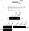
IRBIT is recruited to an exogenous polyadenylation signal-containing RNA in vivo. A, diagram of SV40 late polyadenylation signal used in RNA immunoprecipitation experiments. The cleavage site, USEs, and hexanucleotide (HEX) are represented. The arrow denotes the position of the primer used for reverse transcription that detected both cleaved and uncleaved RNA products. The sequences of wild type (WT) or mutated plasmids used in C are shown, with the USEs and hexanucleotide boxed and the mutations underlined. The USEs were defined according to Ref. . B, HeLa cells were transfected with a plasmid allowing the transcription of SV40 late polyadenylation signal and subjected to immunoprecipitation with anti-IRBIT, anti-CPSF100, or control antibody in the presence (RNase(+)) or the absence (RNase(-)) of RNase A. The RNA was extracted from the immune complex and analyzed by RT-PCR. NT, nontransfected control; -RT, transfected control without reverse transcriptase; M, DNA marker. C, HeLa cells were transfected with plasmids allowing the transcription of wild type or mutated (mUSE, mHEX, or mUSEmHEX) SV40 late polyadenylation signal and processed for RNA immunoprecipitation. The glyceraldehyde-3-phosphate dehydrogenase signal was amplified from reverse transcription products obtained with primer (T)16, to confirm that equivalent amounts of total RNA were present in the input samples.
FIGURE 3.
FIGURE 3.
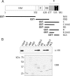
IRBIT binds to the arginine-rich domain of Fip1. A, diagram of mouse Fip1 primary structure and deletion mutants used in B. The numbers indicate amino acid residues. N terminus and middle domain (NM), proline-rich domain (P), mixed charged domain (RD), arginine-rich domain (R), and NLS were assigned according to Ref. . B, COS-7 cells were transfected with HA-IRBIT and processed for pulldown assay with recombinant GST fusion proteins of Fip1 deletion mutants. Pulled down proteins were subjected to Western blotting with anti-HA antibody and to Coomassie Brilliant Blue staining (CBB).
FIGURE 4.
FIGURE 4.
Phosphorylation of the serine-rich region of IRBIT is necessary, but not sufficient, for interaction with Fip1. A, schematic representation of the structure of IRBIT, showing the N-terminal region (NTR), the C-terminal region (CTR), the serine-rich region (SER), and the HA-tagged deletion mutants. B, COS-7 cells were transfected with HA-tagged deletion mutants of IRBIT and processed for pulldown assay with GST or R domain of Fip1. Cell lysates (Input) and pulled down samples were analyzed by Western blotting with anti-HA antibody. C, COS-7 cells were transfected with HA-tagged serine/threonine-substituted mutants of IRBIT and subjected to pulldown assay with GST or R domain of Fip1 and to Western blotting with anti-HA antibody. D, pulldown experiment with recombinant IRBIT purified from E. coli and Sf9 cells. Sf9-expressed IRBIT was incubated with or without alkaline phosphatase (AP) prior to pulldown assay with GST or R domain of Fip1. The samples were analyzed by Western blotting with anti-IRBIT antibody.
FIGURE 5.
FIGURE 5.
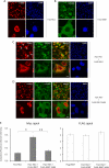
IRBIT promotes the cytoplasmic redistribution of Fip1. AD, COS-7 cells were transfected with Myc-tagged Fip1 (A), FLAG-tagged IRBIT (B), Myc-tagged Fip1 and FLAG-tagged IRBIT (C), and Myc-tagged Fip1 and FLAG-tagged IRBIT-S68A (D) and subjected to immunofluorescence with anti-FLAG and anti-Myc antibodies. The arrows indicate examples of cells showing strong Myc signal in the cytoplasm. Bars, 10 μm. E, the intensity of the signal observed in the cytoplasm and nucleus of Myc-positive cells (left panel) and FLAG-positive cells (right panel) was quantified using National Institutes of Health ImageJ software. The cytoplasm/nucleus signal ratio was calculated for each cell. The values represent means ± S.E. of 170 measures. *, the cytoplasm/nucleus signal ratio of Fip1-Myc expressing cells was significantly higher when cotransfected with FLAG-IRBIT (Student's test; p < 0.01, compared with cells transfected with Fip1-Myc alone). **, the cytoplasm/nucleus signal ratio of Fip1-Myc expressing cells was significantly higher when cotransfected with FLAG-IRBIT than with FLAG-IRBIT-S68A (Student's test; p < 0.01).
FIGURE 6.
FIGURE 6.
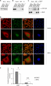
tBHQ treatment modulates the phosphorylation state of endogenous IRBIT and enhances the interaction between IRBIT and CPSF in vitro and in vivo. A, HEK293 cells were treated with 100 μm tBHQ or vehicle (dimethyl sulfoxide (DMSO)) during 4 h and lysed. The lysates were incubated with or without alkaline phosphatase (AP) and analyzed by Western blotting with anti-IRBIT antibody. B, HEK293 cells were treated with tBHQ or vehicle and processed for pulldown assay with GST or R domain of Fip1. The samples were analyzed by Western blotting with anti-IRBIT antibody. C, HEK293 cells were treated with tBHQ or vehicle and subjected to immunoprecipitation with anti-IRBIT or control antibody. The samples were analyzed by Western blotting with anti-CPSF100 and anti-IRBIT antibodies. For IRBIT Western blotting, 10-fold less material was applied in IgG and α-IRBIT lanes than in input sample. D, HeLa cells were treated with tBHQ or vehicle and processed for immunofluorescence with anti-IRBIT and anti-Fip1 antibodies. Bars, 20 μm. E, the intensity of the signal observed in the cytoplasm and nucleus of controls or tBHQ-treated cells was quantified using National Institutes of Health ImageJ software. The cytoplasm/nucleus signal ratio was calculated for each cell. The values represent means ± S.E. of 50 measures. *, the cytoplasm/nucleus ratio of IRBIT signal was significantly lower in tBHQ-treated cells than in controls (Student's test; p < 0.01). **, the cytoplasm/nucleus ratio of Fip1 signal was significantly higher in tBHQ-treated cells than in controls (Student's test; p < 0.01).
FIGURE 7.
FIGURE 7.
IRBIT, Fip1, and PAP form a ternary complex in vitro. A, Sf9-expressed IRBIT, Fip1-Myc and Fip1ΔR-Myc were mixed in various combinations and subjected to pulldown with full-length PAP fused to GST, or GST alone. The input and pulled down samples were analyzed by Western blotting with anti-IRBIT and anti-Myc antibodies and by CBB staining. Open and filled arrowheads indicate the migration of GST and full-length PAP, respectively. B, diagram of PAP primary structure and deletion mutants used in C. The numbers indicate amino acid residues. The catalytic domain (CAT), the RNA-binding domain (RBD), and the C terminus were assigned according to Ref. . The Fip1-binding domain is shown (11). C, Sf9 IRBIT was incubated with GST fusion proteins of PAP deletion mutants. The pulled down proteins were analyzed by Western blotting with anti-IRBIT antibody and by CBB staining.
FIGURE 8.
FIGURE 8.
IRBIT inhibits the polyadenylation activity of PAP in a phosphorylation-dependent manner. A and B, Sf9 IRBIT was treated with 0 (AP(-)), 10 (AP(mild)), or 100 (AP(strong)) units of alkaline phosphatase, purified, and analyzed by CBB staining (A) and Western blotting with anti-phospho-68/71, anti-phospho-74/77, and anti-IRBIT antibodies (B). C, AP(-), AP(mild), or AP(strong) IRBIT were subjected to pulldown assay with GST and full-length PAP. The pulled down proteins were analyzed by Western blotting with anti-IRBIT antibody. D, polyadenylation assays were carried out with internally labeled SV40 late polyadenylation signal RNA (40 fmol) and recombinant PAP (0.1 pmol), in the presence of 0 pmol (lanes 1, 5, 9, 13, and 17), 0.1 pmol (lanes 2, 6, 10, 14, and 18), 0.25 pmol (lanes 3, 7, 11, 15, and 19), and 0.6 pmol (lanes 4, 8, 12, 16, and 20) of AP(-), AP(mild), and AP(strong) IRBIT proteins or the unrelated GST protein (control). Lane M, RNA marker. E, the length of the poly(A) tail was measured on the autoradiogram and plotted against IRBIT amount. The results obtained from one representative experiment are shown. nt, nucleotides.

Similar articles

Cited by

References

    1. Ando, H., Mizutani, A., Matsu-ura, T., and Mikoshiba, K. (2003) J. Biol. Chem. 278 10602-10612 - PubMed
    1. Ando, H., Mizutani, A., Kiefer, H., Tsuzurugi, D., Michikawa, T., and Mikoshiba, K. (2006) Mol. Cell 22 795-806 - PubMed
    1. Shirakabe, K., Priori, G., Yamada, H., Ando, H., Horita, S., Fujita, T., Fujimoto, I., Mizutani, A., Seki, G., and Mikoshiba, K. (2006) Proc. Natl. Acad. Sci. U. S. A. 103 9542-9547 - PMC - PubMed
    1. Mandel, C. R., Bai, Y., and Tong, L. (2008) Cell Mol. Life Sci. 65 1099-1122 - PMC - PubMed
    1. Ryan, K., Calvo, O., and Manley, J. L. (2004) RNA 10 565-573 - PMC - PubMed

Publication types

MeSH terms