. 2009 May;83(9):4308-15.
doi: 10.1128/JVI.02196-08. Epub 2009 Feb 18.

Role of defective Oct-2 and OCA-B expression in immunoglobulin production and Kaposi's sarcoma-associated herpesvirus lytic reactivation in primary effusion lymphoma

Affiliations

Role of defective Oct-2 and OCA-B expression in immunoglobulin production and Kaposi's sarcoma-associated herpesvirus lytic reactivation in primary effusion lymphoma

Daniel L Di Bartolo et al. J Virol. 2009 May.

Abstract

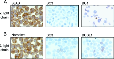
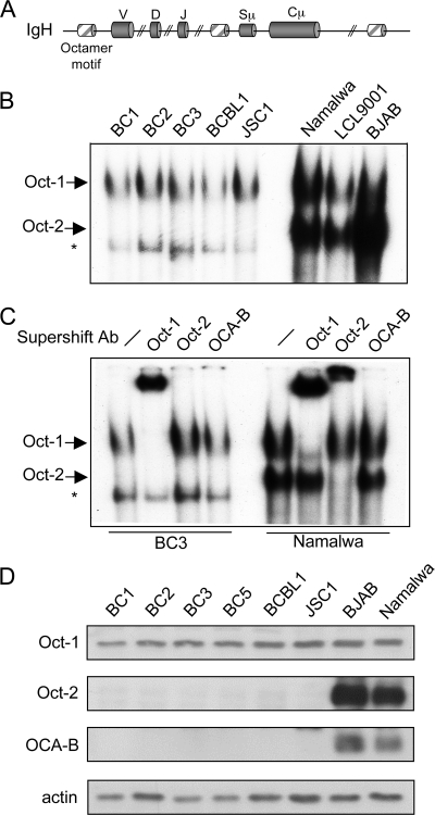
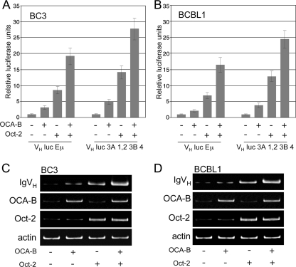
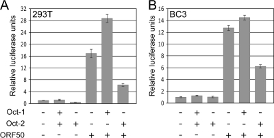
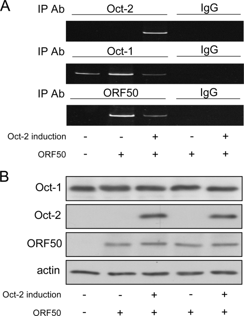
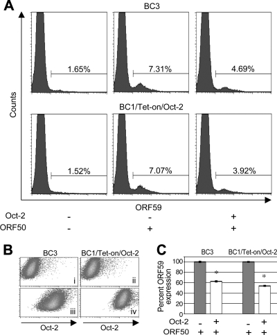
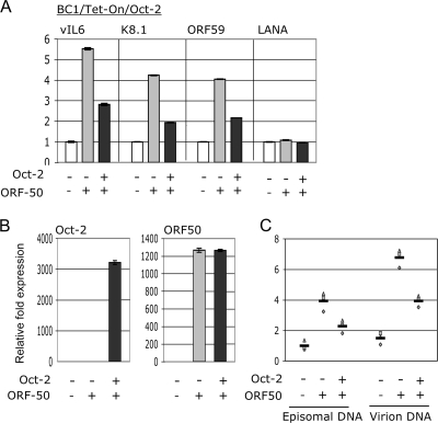
Primary effusion lymphoma (PEL) is a distinct type of B-cell non-Hodgkin lymphoma characterized by the presence of Kaposi's sarcoma-associated herpesvirus (KSHV/human herpesvirus 8). Despite having a genotype and gene expression signature of highly differentiated B cells, PEL does not usually express surface or cytoplasmic immunoglobulin (Ig). We show the lack of Oct-2 and OCA-B transcription factors to be responsible, at least in part, for this defect in Ig production. Like Ig genes, ORF50, the key regulator of the switch from latency to lytic reactivation, contains an octamer motif within its promoter. We therefore examined the impact of Oct-2 and OCA-B on ORF50 activation. The binding of Oct-1 to the ORF50 promoter has been shown to significantly enhance ORF50 transactivation. We found that Oct-2, on the other hand, inhibited ORF50 expression and consequently lytic reactivation by competing with Oct-1 for the octamer motif in the ORF50 promoter. Our data suggest that Oct-2 downregulation in infected cells would be favorable to KSHV in allowing for efficient viral reactivation.


Figures

FIG. 1.
FIG. 1.
PEL cells express low levels of cIg. Cytospins of PEL cell lines were analyzed for expression of κ (A) and λ (B) light chains by immunohistochemistry. BC1 showed very faint κ expression. BC3 was negative for κ light chains but showed rare λ-positive cells. A few BCBL1 cells were positive for λ light chains. BJAB and Namalwa cells were used as positive controls for κ and λ light chain expression, respectively. Experiments were performed at least three independent times.
FIG. 2.
FIG. 2.
PEL cells lack expression of Oct-2 and OCA-B. (A) Schematic representation of octamer motifs found in the Ig(H) locus. Sμ is the switch μ region and Cμ is the constant μ region of Ig(H). (B) Electrophoretic mobility shift assay analysis of the PEL cell lines BC1, BC2, BC3, BCBL1, and JSC1 and the control B-cell lymphoma cell lines Namalwa, LCL9001, and BJAB using an oligonucleotide probe specific for octamer binding proteins. (C) Supershift analysis revealing the makeup of each protein complex. Antibodies to Oct-1, Oct-2, and OCA-B were used. The Oct-1- and Oct-2-containing complexes are labeled; the asterisk indicates a nonspecific complex. (D) Immunoblot analysis of Oct-1, Oct-2, and OCA-B in PEL cells. The Burkitt lymphoma cell lines BJAB and Namalwa were used as positive controls. Data are representative of triplicate experiments.
FIG. 3.
FIG. 3.
IgVH gene expression increases upon ectopic expression of Oct-2 and OCA-B. (A and B) IgVH luciferase reporter activity in BC3 (A) and BCBL1 (B) cells transfected with Oct-2 and/or OCA-B. Both the VH luc Eμ and the VH luc 3A 1,2 3B 4 reporter construct, described in Materials and Methods, were used. Data, representative of three independent experiments, are shown as means of triplicate values ± standard errors of the means. (C and D) Expression of endogenous IgVH transcripts in BC3 (C) and BCBL1 (D) cells, transfected as indicated. IgVH expression was determined by seminested PCR. Expression of Oct-2 and OCA-B was verified by RT-PCR. Data are representative of three independent experiments.
FIG. 4.
FIG. 4.
Oct-2 represses ORF50 transactivation. Shown is pRpORF50 luciferase reporter activity in 293T (A) and BC3 cells (B) transfected as indicated. Experiments were performed independently in triplicate, and representative data are shown as means ± standard errors of the means.
FIG. 5.
FIG. 5.
Oct-2 represses the binding of Oct-1 and ORF50 to the ORF50 promoter. (A) ChIP assay results show the binding of Oct-1, Oct-2, and ORF50 to the endogenous ORF50 promoter in BC1/Tet-on/Oct-2 cells under specified conditions. A control IgG corresponding to each IP antibody was used to verify specificity. Data shown are representative of three independent experiments. (B) Expression of Oct-1, Oct-2, and ORF50 under the various conditions was confirmed by Western blotting.
FIG. 6.
FIG. 6.
Oct-2 represses lytic reactivation. (A) BC3 and BC1/Tet-on/Oct-2 cells were analyzed for ORF59 expression by flow cytometry under untreated conditions (left), upon ORF50 transfection (middle), and upon coexpression of ORF50 and Oct-2 (right). Cells were cotransfected with GFP, and GFP-positive cells were gated to demarcate the transfected population. (B) Expression of Oct-2 in GFP-positive untransfected BC3 cells (i), uninduced BC1/Tet-on/Oct-2 cells (ii), Oct-2-transfected BC3 cells (iii), and (iv) induced BC1/Tet-on/Oct-2 cells. (C) Graph showing reduction in ORF59 expression in ORF50-transfected cells upon Oct-2 expression. All experiments were performed three independent times. P values of ≤0.01, according to Student's t test, were considered significant (*).
FIG. 7.
FIG. 7.
Oct-2 represses lytic replication. (A) qRT-PCR analysis of vIL6, K8.1, ORF59, and LANA in BC1/Tet-on/Oct-2 cells under the specified conditions. (B) qRT-PCR analysis of Oct-2 and ORF50 expression. (C) Analysis of KSHV episomal and encapsidated genomic DNA by real-time PCR. For panels A and B, data are reported as means of triplicate values ± standard errors of the means, and representative data from three independent experiments are shown. In panel C, data from three independent experiments are graphed.

Similar articles

Cited by

References

    1. Annweiler, A., S. Zwilling, R. A. Hipskind, and T. Wirth. 1993. Analysis of transcriptional stimulation by recombinant Oct proteins in a cell-free system. J. Biol. Chem. 2682525-2534. - PubMed
    1. Ansari, M. Q., D. B. Dawson, R. Nador, C. Rutherford, N. R. Schneider, M. J. Latimer, L. Picker, D. M. Knowles, and R. W. McKenna. 1996. Primary body cavity-based AIDS-related lymphomas. Am. J. Clin. Pathol. 105221-229. - PubMed
    1. Arguello, M., M. Sgarbanti, E. Hernandez, Y. Mamane, S. Sharma, M. Servant, R. Lin, and J. Hiscott. 2003. Disruption of the B-cell specific transcriptional program in HHV-8 associated primary effusion lymphoma cell lines. Oncogene 22964-973. - PubMed
    1. Babb, R., M. A. Cleary, and W. Herr. 1997. OCA-B is a functional analog of VP16 but targets a separate surface of the Oct-1 POU domain. Mol. Cell. Biol. 177295-7305. (Erratum, 18:2430, 1998.) - PMC - PubMed
    1. Banerji, J., L. Olson, and W. Schaffner. 1983. A lymphocyte-specific cellular enhancer is located downstream of the joining region in immunoglobulin heavy chain genes. Cell 33729-740. - PubMed

Publication types

MeSH terms

LinkOut - more resources