. 2009 May;83(9):4508-19.
doi: 10.1128/JVI.02429-08. Epub 2009 Feb 18.

The marburg virus 3' noncoding region structurally and functionally differs from that of ebola virus

Affiliations

The marburg virus 3' noncoding region structurally and functionally differs from that of ebola virus

Sven Enterlein et al. J Virol. 2009 May.

Abstract

We have previously shown that the first transcription start signal (TSS) of Zaire Ebola virus (ZEBOV) is involved in formation of an RNA secondary structure regulating VP30-dependent transcription activation. Interestingly, transcription of Marburg virus (MARV) minigenomes occurs independently of VP30. In this study, we analyzed the structure of the MARV 3' noncoding region and its influence on VP30 necessity. Secondary structure formation of the TSS of the first gene was experimentally determined and showed substantial differences from the structure formed by the ZEBOV TSS. Chimeric MARV minigenomes mimicking the ZEBOV-specific RNA secondary structure were neither transcribed nor replicated. Mapping of the MARV genomic replication promoter revealed that the region homologous to the sequence involved in formation of the regulatory ZEBOV RNA structure is part of the MARV promoter. The MARV promoter is contained within the first 70 nucleotides of the genome and consists of two elements separated by a spacer region, comprising the TSS of the first gene. Mutations within the spacer abolished transcription activity and led to increased replication, indicating competitive transcription and replication initiation. The second promoter element is located within the nontranslated region of the first gene and consists of a stretch of three UN(5) hexamers. Recombinant full-length MARV clones, in which the three conserved U residues were substituted, could not be rescued, underlining the importance of the UN(5) hexamers for replication activity. Our data suggest that differences in the structure of the genomic replication promoters might account for the different transcription strategies of Marburg and Ebola viruses.


Figures

FIG. 1.
FIG. 1.
Sequence comparison of the 3′ NCR of MARV Musoke (GenBank accession number DQ217792), ZEBOV (GenBank accession number NC_002549), Reston ebolavirus (REBOV; GenBank accession number AY769362), and Sudan ebolavirus (SEBOV; GenBank accession number NC_006432). The sequences were aligned to maximize stretches of similarity. The transcription start signal of the first gene (NP) is underlined. Uridine residues that appear every 6 nt in at least three consecutive hexamers and adjacent purine residues are in boldface. In an alternative frame, these residues are marked with a circle (Ů).
FIG. 2.
FIG. 2.
Results of the chemical modification assay on RNA of the positive-strand minigenome 3M-5MG(+). In vitro-transcribed RNA was subjected to treatment with either DMS or CMCT to specifically modify A and C or G and U residues, respectively. The modified RNAs were used as the template in an RT reaction with a 32P-labeled primer, and the products were separated on a denaturing polyacrylamide gel. Modified bases led to termination of RT. To relate the pattern of the RNA (left four lanes) to the template, plasmid DNA was sequenced using the same primer and run along with the RT product (right four lanes). −, RT products of the untreated RNA template as a background control. (A) Modification pattern of the first 48 nt of the RNA comprising the leader region. Nucleotides that were modified are indicated and are shaded in gray in the sequence below. (B) Modification pattern from the transcription start signal (nt 49 to 60, boxed) to nt 102. Modified nucleotides are marked as described for panel A. The transcription start signal of NP gene is boxed. (C) Interpretation of the modification pattern. A model of the secondary structure was predicted using the online application Mfold; this model was then adapted according to the results of the chemical modification assay. Sequence that was not interpretable is shown in gray. Modified nucleotides are underlined, and asterisks mark the base pairs of which only one base was modified. The transcription start signal is marked by a line. (D) The secondary structure of the ZEBOV transcription start signal as a comparison. The transcription start signal itself is marked by a line.
FIG. 3.
FIG. 3.
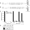
Chimeric minigenomes between ZEBOV and MARV transcription start signals. (A) Mutational changes to replace the MARV NP transcription start signal with that of ZEBOV, including the downstream sequence involved in formation of a stable hairpin structure. Mutated nucleotides are shown in boldface and are marked by asterisks. The predicted RNA secondary structure is shown on the right. (B) Changes introduced to resemble the ZEBOV-specific hairpin structure (scheme at right) but keep the MARV-specific transcription start signal. The transcription start signal is underlined for both sequences, and the exchanged nucleotides are marked with asterisks. (C) The two chimeric constructs were tested with the minigenome assay. BSR-T7/5 cells were transfected with either ZEBOV-specific expression plasmids encoding the nucleocapsid proteins NP (0.5 μg), VP35 (0.5 μg), L (1.0 μg), and VP30 (0.1 μg; left part) or plasmids encoding MARV nucleocapsid proteins (NP, 0.1 μg; VP35, 0.5 μg; L, 1.0 μg; and VP30, 0.5 μg; right part); the wild-type (wt) minigenome was 1.0 μg 3E-5E and 3M-5M, respectively. Plasmids for VP30 and L were added as indicated. At 2 days posttransfection, cells were harvested and CAT activity was determined. Positive controls were set as 100%. VP30E, ZEBOV VP30; VP30M, MARV VP30. (D) Analysis of replicated RNA by Northern blot analysis. Huh-T7 cells were transfected with all necessary MARV minigenome plasmids. As the negative control, the plasmid encoding the L gene was omitted. At 2 days posttransfection, cells were lysed and treated with micrococcal nuclease. Protected RNA was purified and subjected to Northern blot analysis. M→E, MARV→EBOV; M1→E2, MARV1→EBOV2.
FIG. 4.
FIG. 4.
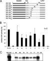
Mapping of the MARV 3′ NCR. (A) Schematic drawing of the 3′ end of the MARV minigenomic deletion mutants. Nucleotides are numbered according to their position in the viral RNA. Deleted sequence is indicated by a dashed line. tss, transcription start signal; CAT, CAT gene. (B) Huh-T7 cells were transfected with plasmids encoding MARV NP, VP35, and L along with the respective truncated minigenome. At 2 days posttransfection, cells were lysed and tested for CAT activity, reflecting replication and transcription of the minigenomes. The positive control (3M-5M) was set as 100%. The experiment was performed three times, and the standard deviations are shown. (C) Northern blot analysis showing replicated RNA of the deletion mutants. Numbers indicate the lengths (in nucleotides) of the 3′ ends of the truncated minigenomes.
FIG. 5.
FIG. 5.
Characterization of the MARV genomic replication promoter. Various point mutations were introduced into the 3′ NCR of 3M-5M. Huh-T7 cells were transfected with the minigenome system components and analyzed for replication and transcription at 48 h posttransfection. CAT activity reflects transcription activity, and Northern blot analysis was performed to test for replicated RNA. (A) CAT assay (left) and Northern blot analysis (right) of minigenomes with point mutations in a region of two identical adjacent hexamers located downstream of the transcription start signal. (B) CAT assay (left) and Northern blot analysis (right) of minigenomes in which various hexameric U residues in the 3′ NCR of the NP gene were replaced with A. Mutated nucleotides are indicated.
FIG. 6.
FIG. 6.
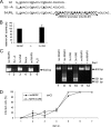
Rescue of recombinant MARV. (A) MARV mutants. U residues 62, 68, and 74 were replaced with A in the full-length MARV mutant 3U→A. The full-length mutant 3 × UN5 contains three extra hexamers derived from the ZEBOV replication promoter. Hexameric U residues are in boldface and underlined. (B) CAT assay of MARV minigenome containing three additional EBOV UN5 hexamers. BSR-T7/5 cells were transfected as previously described and analyzed for CAT expression after 48 h. (C) RT-PCR to detect recovered recombinant MARV. BSR-T7/5 cells were transfected with plasmids encoding MARV NP, VP35, VP30, and L and the respective full-length clone along with pC-T7/Pol. At 5 days posttransfection, transfected cells were mixed with fresh Vero C1008 cells and cocultivated for another 6 days. Cells were then lysed, and the lysates were used to infect fresh Vero C1008 cells. Infected cells were lysed at 7 days postinfection, and cellular RNA was used for RT-PCR to amplify nt 5890 to 6521 of the MARV genome. In contrast to the wild-type virus, recombinant MARV viruses contain an additional SspI restriction site within the amplified fragment. The 632-bp fragment was digested with SspI where noted. Two bands, at 301 and 331 bp, indicate the presence of the SspI site. rec MARV, recombinant MARV containing the additional SspI site as a genetic tag; Mock, not transfected and not infected; wt MARV, infected with wild-type MARV. (D) Growth characteristics of recombinant MARV and MARV3xUN5. Huh7 cells were infected with either recombinant MARV or MARV3xUN5 at an MOI of 0.01. Cells were harvested daily up to day 7 and subjected to immunofluorescence analysis using a MARV-specific antiserum. Cell nuclei were stained with DAPI. Infectivity of the viruses was determined by counting fluorescent cells. Data were obtained in triplicate, and the standard deviations are shown.
FIG. 7.
FIG. 7.
Mutations within the transcription start signal of the NP gene led to enhanced replication. Point mutations were introduced into the transcription start signal and downstream-located sequences of minigenome 3M-5M. The mutation sites are indicated. Huh-T7 cells were transfected with plasmids encoding the MARV nucleocapsid proteins and the respective mutated minigenome. At 48 h posttransfection, cells were lysed and subjected to a CAT assay, reflecting transcription activity, and Northern blot analysis to show replication activity. (A) CAT assay (left) and Northern blot analysis (right) of minigenomes containing mutations within the transcription start signal of the NP gene. (B) Northern blot analysis of minigenomes in which hexameric U residues located within the transcription start signal of the NP gene and adjacent nucleotides are replaced with A.
FIG. 8.
FIG. 8.
Comparison of the genomic replication promoter of ZEBOV and MARV. Promoter elements (PE) for replication are represented by black boxes, the transcription start signal (TSS) of the NP gene is shown in gray, and additional sequences belonging to the spacer region are indicated in a lighter gray. Regions of unimportant sequence are shown with hatching. Nucleotides involved in secondary structure formation are boxed in the sequence below the scheme. Right insets, scheme of predicted RNA secondary structures of these regions. The NP transcription start signal is indicated by a solid line in the insets and is underlined in the sequence. (A) The ZEBOV replication promoter is bipartite (PE1 and PE2). The spacer region includes the transcription start signal as well as the downstream sequence involved in secondary structure formation and can be extended or reduced by a multiple of 6 nt. PE2 consists of eight hexamers with U residues (boldface) at positions 81, 87,…123. (B) 3′ end of the MARV genome. PE2 of the MARV replication promoter is shorter, containing 3 UN5 hexamers. The spacer region between PE1 and PE2 consists of the transcription start signal of the NP gene. ORF, open reading frame.

Similar articles

Cited by

References

    1. Buchholz, U. J., S. Biacchesi, Q. N. Pham, K. C. Tran, L. Yang, C. L. Luongo, M. H. Skiadopoulos, B. R. Murphy, and P. L. Collins. 2005. Deletion of M2 gene open reading frames 1 and 2 of human metapneumovirus: effects on RNA synthesis, attenuation, and immunogenicity. J. Virol. 796588-6597. - PMC - PubMed
    1. Buchholz, U. J., S. Finke, and K. K. Conzelmann. 1999. Generation of bovine respiratory syncytial virus (BRSV) from cDNA: BRSV NS2 is not essential for virus replication in tissue culture, and the human RSV leader region acts as a functional BRSV genome promoter. J. Virol. 73251-259. - PMC - PubMed
    1. Calain, P., M. C. Monroe, and S. T. Nichol. 1999. Ebola virus defective interfering particles and persistent infection. Virology 262114-128. - PubMed
    1. Calain, P., and L. Roux. 1993. The rule of six, a basic feature for efficient replication of Sendai virus defective interfering RNA. J. Virol. 674822-4830. - PMC - PubMed
    1. Collins, P. L., E. Camargo, and M. G. Hill. 1999. Support plasmids and support proteins required for recovery of recombinant respiratory syncytial virus. Virology 259251-255. - PubMed

Publication types

MeSH terms