. 2009 Mar 10;106(10):4036-41.
doi: 10.1073/pnas.0811277106. Epub 2009 Feb 18.

K+-dependent paradoxical membrane depolarization and Na+ overload, major and reversible contributors to weakness by ion channel leaks

Affiliations

K+-dependent paradoxical membrane depolarization and Na+ overload, major and reversible contributors to weakness by ion channel leaks

Karin Jurkat-Rott et al. Proc Natl Acad Sci U S A. .

Abstract

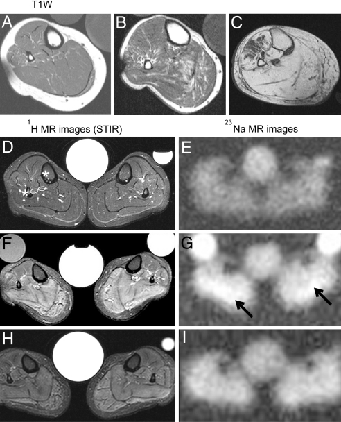
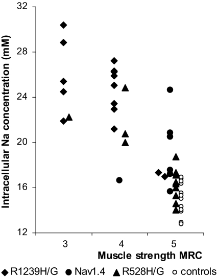
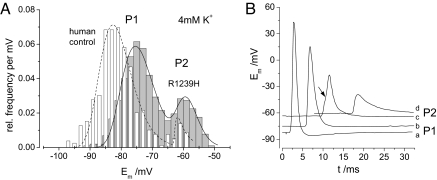
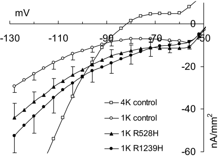
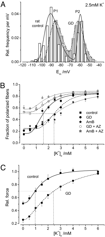
Normal resting potential (P1) of myofibers follows the Nernst equation, exhibiting about -85 mV at a normal extracellular K(+) concentration ([K(+)](o)) of 4 mM. Hyperpolarization occurs with decreased [K(+)](o), although at [K(+)](o) < 1.0 mM, myofibers paradoxically depolarize to a second stable potential of -60 mV (P2). In rat myofiber bundles, P2 also was found at more physiological [K(+)](o) and was associated with inexcitability. To increase the relative frequency of P2 to 50%, [K(+)](o) needed to be lowered to 1.5 mM. In the presence of the ionophore gramicidin, [K(+)](o) reduction to only 2.5 mM yielded the same effect. Acetazolamide normalized this increased frequency of P2 fibers. The findings mimic hypokalemic periodic paralysis (HypoPP), a channelopathy characterized by hypokalemia-induced weakness. Of myofibers from 7 HypoPP patients, up to 25% were in P2 at a [K(+)](o) of 4 mM, in accordance with their permanent weakness, and up to 99% were in P2 at a [K(+)](o) of 1.5 mM, in accordance with their paralytic attacks. Of 36 HypoPP patients, 25 had permanent weakness and myoplasmic intracellular Na(+) ([Na(+)](i)) overload (up to 24 mM) as shown by in vivo (23)Na-MRI. Acetazolamide normalized [Na(+)](i) and increased muscle strength. HypoPP myofibers showed a nonselective cation leak of 12-19.5 microS/cm(2), which may explain the Na(+) overload. The leak sensitizes myofibers to reduced serum K(+), and the resulting membrane depolarization causes the weakness. We postulate that the principle of paradoxical depolarization and loss of function upon [K(+)](o) reduction may apply to other tissues, such as heart or brain, when they become leaky (e.g., because of ischemia).


Conflict of interest statement

The authors declare no conflict of interest.

Figures

Fig. 1.
Fig. 1.
1H and 23Na measurements in the calf muscles of HypoPP patients. (A–C) Axial T1-weighted MR images from 3 related Cav1.1-R1239H patients: a 35-year-old woman (A), her 55-year-old uncle (B), and her 80-year-old grandmother whose limb muscles were predominantly replaced with fat (C). (D–I) T2-weighted STIR 1H (Left) and 23Na-MR images (Right) from a healthy control (D and E) and a 37-year-old patient (F–I), the sister of the patient in A. The images in F and G were taken before treatment, and the images in H and I were taken after treatment with 250 mg/d AZ for 4 weeks. Note the very high hydrogen intensities in the STIR sequence (F) and the elevated Na+ concentration before treatment (G, arrows indicate highest Na signal intensities) and their improvement after treatment. The central reference contains 0.3% NaCl solution; occasional side tubes containing 0.3% NaCl in 1% agarose (Left) and 0.6% NaCl in H2O (Right) were additional standards.
Fig. 2.
Fig. 2.
Correlation of strength and [Na+]i in 36 HypoPP patients. The [Na+]i values obtained with a 23Na-MRI of the lower legs and the values of plantar flexor muscle strength measured according to the MRC grading scale (closed symbols) show a clear correlation. Values from 12 healthy controls are shown as open circles.
Fig. 3.
Fig. 3.
Resting (Em) and action potentials of normal and HypoPP fibers. (A) Bimodal distribution of Em of human control fibers (white, n = 369) and R1239H-HypoPP muscle fibers (gray, n = 128) in 4-mM K+. HypoPP fibers have an increased probability for the second stable membrane potential of −60 mV (P2); the first stable membrane potential (P1) in HypoPP fibers is depolarized by ≈ 10 mV compared with the controls. (B) Action potentials (AP) in the endplate region elicited from various holding potentials of −82 mV (a), −75 mV (b), −62 mV (c), and −59 mV (d). At c there is a minimal AP beginning at the arrow; at d there is no AP, only the endplate potential.
Fig. 4.
Fig. 4.
Kir conductance and cation leak in human myofibers. Steady-state current density–voltage relationships were determined in 1.0-mM K+ for 7 normal myofibers (1K control) and 4 (1K R528H) and 5 myofibers (1K R1239H) from patients. The difference between the curves for patients and controls corresponds to the mutation-related leak current. In addition, 8 normal myofibers (4K control) were measured in 4.0-mM K+. Note that the 4K curve is much steeper (gmax = 261 μS/cm2 at hyperpolarization) than the 1K curve (gmax = 26.5 μS/cm2) of the control fibers according to the higher Kir conductance in 4-mM K+. To avoid superimposing currents, the solutions contained TTX, not Cl.
Fig. 5.
Fig. 5.
Resting potentials (Em) of rat muscle strips treated with GD. (A) Bimodal distribution of Em in control (white, n = 257 fibers) and rat diaphragm strips treated with 0.1 μM GD (gray, n = 265 fibers) to create leaks. As in HypoPP, the relative frequency of the second stable potential (P2) at −60 mV increased, and the first stable potential (P1) was depolarized by ≈ 5 mV for the leaky fibers. (B) Relative P1 frequency dependent on external K+. Fitting the data points (6–7 strips each) to a multimodal log-normal probability density function enabled comparison. The turning point is at 2.5 mM K+ in GD-treated leaky fibers, at 2.9 mM in AmB-treated fibers, and at 1.5 mM for controls, suggesting that the leaky fibers have a higher probability for the second stable potential (P2 = −60 mV) when [K+]o decreases. The addition of 100 μM AZ to the leaky fibers resulted in the restoration of the fraction of polarized fibers, particularly in low K+. (C) Dependence of twitch force on [K+]o (5 strips at each data point). The force reduction in GD-treated muscles and control muscles upon [K+]o reduction reflects the loss of polarized fibers illustrated in B.

References

    1. Venance SL, et al. The primary periodic paralyses: Diagnosis, pathogenesis and treatment. Brain. 2006;129:8–17. - PubMed
    1. Links TP, Zwarts MJ, Wilmink JT, Molenaar WM, Oosterhuis HJ. Permanent muscle weakness in familial hypokalaemic periodic paralysis. Clinical, radiological and pathological aspects. Brain. 1990;113:1873–1889. - PubMed
    1. Rüdel R, Lehmann-Horn F, Ricker K, Küther G. Hypokalemic periodic paralysis: In vitro investigation of muscle fiber membrane parameters. Muscle Nerve. 1984;7:110–120. - PubMed
    1. Ruff RL. Insulin acts in hypokalemic periodic paralysis by reducing inward rectifier K+ current. Neurology. 1999;53:1556–1563. - PubMed
    1. Geukes Foppen RJ, van Mil HG, van Heukelom JS. Effects of chloride transport on bistable behaviour of the membrane potential in mouse skeletal muscle. J Physiol. 2002;542:181–191. - PMC - PubMed

MeSH terms