. 2009 Apr;20(8):2311-26.
doi: 10.1091/mbc.e08-12-1169. Epub 2009 Feb 18.

Regulation of distinct septin rings in a single cell by Elm1p and Gin4p kinases

Affiliations

Regulation of distinct septin rings in a single cell by Elm1p and Gin4p kinases

Bradley S DeMay et al. Mol Biol Cell. 2009 Apr.

Abstract

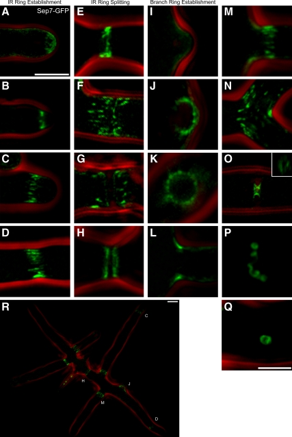
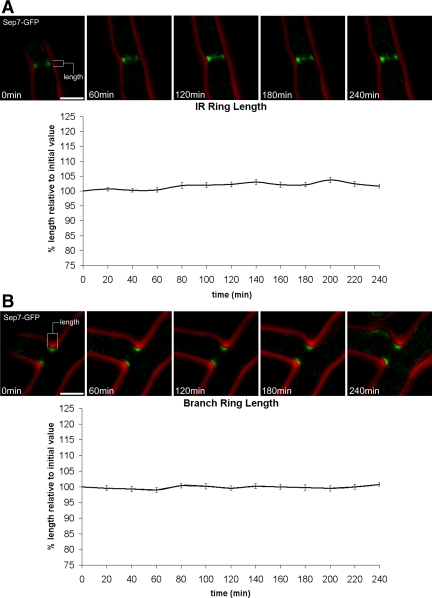
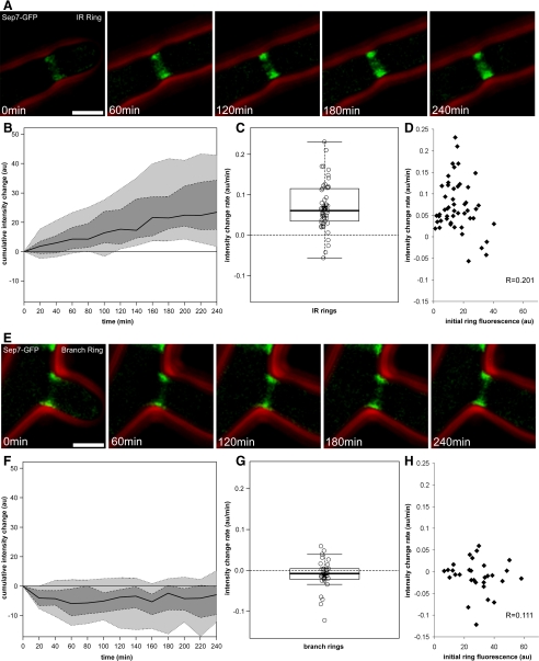
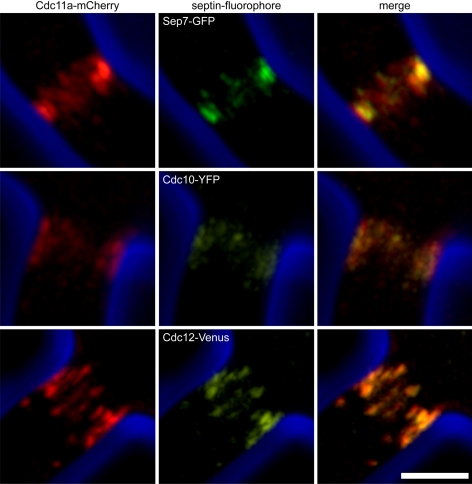
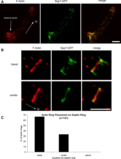
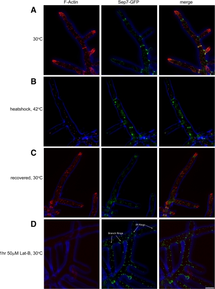
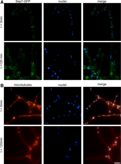
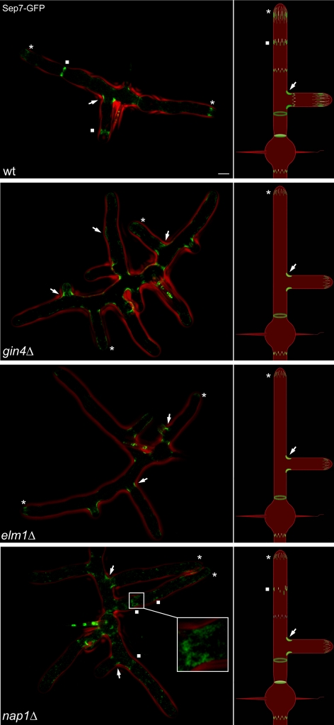
Septins are conserved, GTP-binding proteins that assemble into higher order structures, including filaments and rings with varied cellular functions. Using four-dimensional quantitative fluorescence microscopy of Ashbya gossypii fungal cells, we show that septins can assemble into morphologically distinct classes of rings that vary in dimensions, intensities, and positions within a single cell. Notably, these different classes coexist and persist for extended times, similar in appearance and behavior to septins in mammalian neurons and cultured cells. We demonstrate that new septin proteins can add through time to assembled rings, indicating that septins may continue to polymerize during ring maturation. Different classes of rings do not arise from the presence or absence of specific septin subunits and ring maintenance does not require the actin and microtubule cytoskeletons. Instead, morphological and behavioral differences in the rings require the Elm1p and Gin4p kinases. This work demonstrates that distinct higher order septin structures form within one cell because of the action of specific kinases.


Figures

Figure 1.
Figure 1.
Sep7-GFP localizes to diverse higher order septin structures. Sep7-GFP (AG127)–expressing cells were imaged live after growth overnight on media containing-agarose pad except where noted. All images are maximum projections of image stacks. (A and B) Sep7p at the hyphal tip. (C) A nascent IR ring. (D and E) IR rings. (F–H) Splitting of an IR ring and septation. (I) Sep7p at an incipient branch site. (J) Branch ring. (K) Cross section of branch ring. (L) Branch ring. (M) IR ring formed adjacent to branch ring. (N) IR ring traversing a Y-junction. (O) Hourglass-shaped septin structure at a septum. (P) Septin helix formed under stress. (Q) Cortically associated ∼1-μm ectopic ring. (R) Small mycelia in which some examples of the classes of rings shown in A–Q are noted highlighted by letters. (K and P) Cell was fixed with 2% paraformaldehyde. GFP fluorescence (green) and phase contrast outlines (red) are shown. Bars, 5 μm (A–J and L–O; in A); 2.5 μm (K, P, and Q; in Q); and 5 μm (R).
Figure 2.
Figure 2.
Septin rings do not change dimensions with time. Single SEP7-GFP (AG127) cells were observed using 3D time-lapse fluorescence microscopy. Image stacks were acquired every 20 min. All images were acquired and processed identically. The sequences of images above the graphs are examples of an IR ring (A) and a branch ring (B) that were measured. (A) Individual IR septin rings were measured for 4 h after their establishment. N = 50. (B) Branch rings were measured for 4 h after their establishment. N = 30. The small fluctuations in these measurements reflect minor shifts in the orientation of the hypha containing the ring as it grows. GFP fluorescence (green) and phase contrast cell outlines (red) are shown. Bars, 5 μm.
Figure 3.
Figure 3.
Sep7-GFP intensity increases with time at IR septin rings but not at branch rings. Single SEP7-GFP (AG127) cells were observed using 3D time-lapse fluorescence microscopy. Image stacks were acquired every 20 min and were processed identically. (A and E) Images show examples of an IR ring (A) and a branch ring (E) that were measured. The GFP fluorescence (green) and phase contrast cell outlines (red) are shown. Bars, 5 μm. (B and F) Cumulative change in fluorescence intensity of Sep7-GFP at IR rings (B; N = 50) and branch rings (F; N = 30) was measured for 4 h after establishment. Change in fluorescence intensity was calculated by subtracting the intensity of the ring immediately after establishment (t = 0) from the intensity measured at a given time point. Intensity values were corrected for background fluorescence of the cytosol. Values are given in arbitrary fluorescence units (au). The black lines represent the median cumulative change for the population. Fifty percent of the measured rings are in the dark gray regions, and 80% of the rings fall in the combined light gray and dark gray regions. (C and G) The box and whisker plots show the distribution of slopes of the linear regression of fluorescence change at individual rings. The black line denotes the median value and the asterisk shows the mean value. The box is the middle 50% of the data, and the whiskers extend to the most extreme data point, no more than 1.5 times the interquartile range. Rates are given in arbitrary fluorescence units per minute (au/min). (D and H) The slopes of the linear regression of fluorescence changes for a ring are graphed against the initial fluorescence at that ring for each individual septin ring analyzed. Values are given in arbitrary fluorescence units and rates are given in arbitrary fluorescence units per minute. In both cases, R values are insignificant.
Figure 4.
Figure 4.
All classes of septin rings contain all septin subunits. A. gossypii strains expressing Cdc11a-mCherry from the endogenous locus and either Sep7-GFP (AG232), Cdc10-YFP (AG306), or Cdc12-Venus(AG308) expressed using their endogenous promoter from a replicating plasmid were imaged in live cells grown overnight on agarose media pads. mCherry fluorescence (red), GFP fluorescence (green), Venus fluorescence (yellow), and phase contrast outlines (blue) are shown. Bar, 5 μm.
Figure 5.
Figure 5.
F-Actin rings are asymmetrically localized at septin rings. (A) Colocalization of Sep7-GFP and F-actin. SEP7-GFP cells (AG127) were grown overnight in liquid media at 30°C. Cells were fixed with 2% paraformaldehyde and the F-actin was visualized using AlexaFluor-Phalloidin568. (B) Actin rings localized to the basal end (top) or center (bottom) of established septin rings. Arrows point to the hyphal tip and branch point. GFP fluorescence (green), Alexa568-Phalloidin (red), and phase contrast cell outlines (blue) are shown. Bar is 3 μm. (C) Actin ring position was scored relative to the associated septin ring (N = 141).
Figure 6.
Figure 6.
F-actin is required for septin ring assembly but not maintenance. SEP7-GFP cells (AG127) were grown overnight in liquid AFM at 30°C. (A) Before heat shock, cells were fixed with 2% paraformaldehyde, and the F-actin was visualized using AlexaFluor-Phalloidin568. (B) Cells were transferred to 42°C for 20 min to depolarize the actin cytoskeleton and then processed for F-actin visualization. (C) Heat-shocked cells were returned to 30°C to recover for 20 min and then processed for F-actin visualization. (D) Cells were incubated for 1 h in growth media with latrunculin-B (final concentration, 50 μM) and then processed for F-actin visualization. GFP fluorescence (green), AlexaFluor-Phalloidin568 fluorescence (red), and phase contrast cell outlines (blue) are shown. Bar, 5 μm.
Figure 7.
Figure 7.
Microtubules are not required for septin ring assembly or maintenance. Cells expressing Sep7-GFP (AG127) were grown overnight in liquid AFM media at 30°C (t = 0) and then were treated with 20 μg/ml nocodazole for 120 min and fixed for visualization of Sep7-GFP (A) or anti-α-tubulin immunofluorescence (B) in combination with Hoechst staining of DNA. Sep7-GFP (green), anti-α-tubulin (red), and Hoechst (blue) stainings are shown. Bars, 5 μm.
Figure 8.
Figure 8.
Deletion of septin regulators disrupts IR rings but not branch rings. Live cell fluorescence images of wild-type SEP7-GFP (AG127), gin4Δ SEP7-GFP (AG121), elm1Δ SEP7-GFP (AG120), and nap1Δ SEP7-GFP (AG209) cells grown overnight on agarose pads. Asterisks, squares, and arrows highlight the presence of tip-associated filaments, interregion rings, and branch rings, respectively. In the nap1Δ panel, an asymmetric interregion ring is highlighted and enlarged. GFP fluorescence (green) and phase contrast cell outlines (red) are shown. Bar, 5 μm.
Figure 9.
Figure 9.
nap1Δ mutants assemble asymmetric IR rings. Images from a time-lapse series in which nap1Δ (AG314) cells were grown on an agar gel pad and imaged every 30 min. Sep7-GFP is asymmetrically deposited in IR rings as they form (arrow and arrowhead). Bar, 5 μm.

Similar articles

Cited by

References

    1. Alberti-Segui C., Dietrich F., Altmann-Johl R., Hoepfner D., Philippsen P. Cytoplasmic dynein is required to oppose the force that moves nuclei towards the hyphal tip in the filamentous ascomycete Ashbya gossypii. J. Cell Sci. 2001;114:975–986. - PubMed
    1. Ayad-Durieux Y., Knechtle P., Goff S., Dietrich F., Philippsen P. A PAK-like protein kinase is required for maturation of young hyphae and septation in the filamentous ascomycete Ashbya gossypii. J. Cell Sci. 2000;113:4563–4575. - PubMed
    1. Barral Y., Mermall V., Mooseker M. S., Snyder M. Compartmentalization of the cell cortex by septins is required for maintenance of cell polarity in yeast. Mol. Cell. 2000;5:841–851. - PubMed
    1. Baudin A., Ozier-Kalogeropoulos O., Denouel A., Lacroute F., Cullin C. A simple and efficient method for direct gene deletion in Saccharomyces cerevisiae. Nucleic Acids Res. 1993;21:3329–3330. - PMC - PubMed
    1. Bertin A., McMurray M. A., Grob P., Park S. S., Garcia G., 3rd, Patanwala I., Ng H. L., Alber T., Thorner J., Nogales E. Saccharomyces cerevisiae septins: supramolecular organization of heterooligomers and the mechanism of filament assembly. Proc. Natl. Acad. Sci. USA. 2008;105:8274–8279. - PMC - PubMed

Publication types

LinkOut - more resources