. 2009 Jul;23(7):1320-8.
doi: 10.1038/leu.2009.19. Epub 2009 Feb 19.

Cyclin D1 as a universally expressed mantle cell lymphoma-associated tumor antigen for immunotherapy

Affiliations

Cyclin D1 as a universally expressed mantle cell lymphoma-associated tumor antigen for immunotherapy

M Wang et al. Leukemia. 2009 Jul.

Abstract

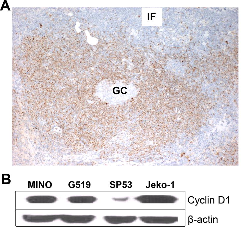
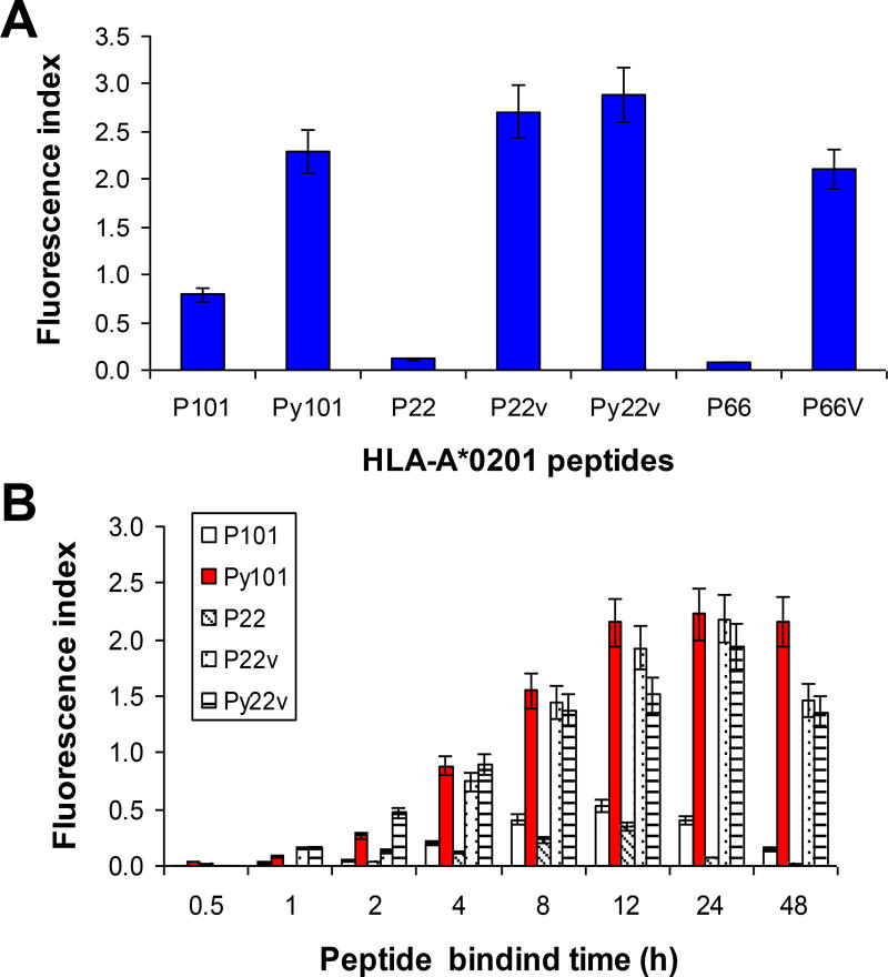
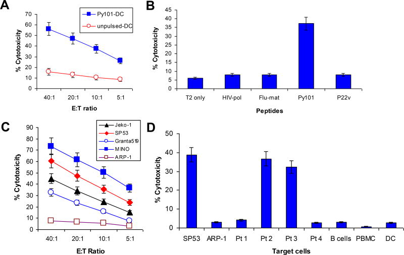
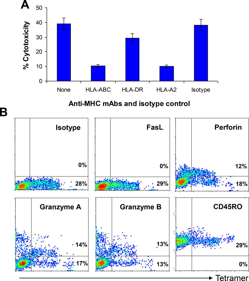
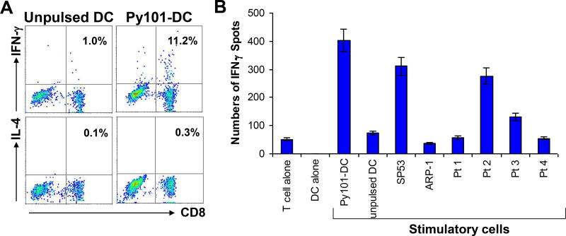
Mantle cell lymphoma (MCL) accounts for 5-10% of all non-Hodgkin lymphomas and has the worst prognosis among all lymphomas. The hallmark of MCL is a t(11;14) translocation that results in overexpression of cyclin D1 by tumor cells of virtually all patients. In this study, we examined whether cyclin D1 could be an effective tumor-associated antigen for immunotherapy. We identified cyclin D1 peptides for HLA-A(*)0201 and generated peptide-specific CD8(+) T-cell lines from HLA-A(*)0201(+) blood donors and MCL patients. These cell lines proliferated in response to cyclin D1 peptide-pulsed stimulatory cells. Moreover, the T cells efficiently lysed peptide-pulsed but not unpulsed T2 cells and autologous dendritic cells; cyclin D1(+) and HLA-A(*)0201(+) human MCL lines MINO, SP53, Jeko-1 and Granta 519; and more importantly, HLA-A(*)0201(+) primary lymphoma cells from MCL patients. No killing was observed with HLA-A(*)0201(-) primary lymphoma cells or HLA-A(*)0201(+) normal blood cells, including B cells. These results indicate that these T cells are potent cytotoxic T cells and recognize cyclin D1 peptides naturally presented by patient lymphoma cells in the context of HLA-A(*)0201 molecules. Taken together, our work identifies cyclin D1 as a potentially important antigen for immunotherapy of MCL.


Figures

Figure 1
Figure 1
Expression of cyclin D1 in primary MCL and established cell lines. (A) Immunohistochemistry staining showing the expression of cyclin D1 by lymphoma cells with a predominantly mantle zone pattern, while normal B cells and other lymph node cells in the germinal center (GC) and the interfollicular area (IF) are negative for cyclin D1. The photomicrographs shown are at ×100 magnification. A representative staining of one lymph node sample from a MCL patient out of 4 examined is shown. (B) Western blots showing cyclin D1 protein expression in HMCLLs MINO, SP53, Jeko-1, and Granta 519 (G519). Representative result of one experiment out of 3 performed is shown.
Figure 2
Figure 2
Binding affinity and stability of cyclin D1 peptides with HLA-A*0201 molecules. Peptide binding assay showing (A) binding affinity and (B) stability (fluorescence index) of two cyclin D1 peptides P101 and P22 and their heteroclitic peptides Py101, P22v, and Py22v for HLA-A*0201 molecules. DKK1 peptide P66 and its heteroclitic peptide P66v were used as controls in (A). In (A) T2 cells were incubated with 100 µg/mL peptides overnight, and in (B) T2 cells were incubated with 100 µg/mL peptides for different time points, and analyzed for surface HLA-A*0201 expression. Details are provided in the Methods section. Representative results of three independent experiments are shown.
Figure 3
Figure 3
Generation of cyclin D1 peptide-specific T-cell lines. (A) Frequency of cyclin D1 Py101 peptide-specific CD8+ T cells measured by HLA-A*0201 peptide-tetramer staining in cultures during in vitro stimulations. Proliferative responses measured by (B) 3[H]-thymidine incorporation or (C) CFSE dilution assays of T cells specific for Py101 peptide in response to autologous Py101 peptide-pulsed (Py101-DC) or unpulsed DCs. Figures inside dot plots or histograms represent the percentages of T cells. Shown are the results of a T-cell line generated from a HLA-A*0201+ blood donor. Similar results are obtained with other Py101 peptide-specific T-cell lines from HLA-A*0201+ blood donors or MCL patients.
Figure 4
Figure 4
Cytolytic activity of cyclin D1 peptide-specific T cells. Shown is the cytotoxicity of cyclin D1 Py101-specific T cells against: (A) Autologous DCs pulsed with or without Py101 peptide; (B) T2 cells pulsed with or without different peptides, including cyclin D1 Py101 and P22v, or control peptides HIV-pol and Flu-matrix (Flu-mat); (C) HMCLLs SP53, MINO, Grant 519, and Jeko-1, and a myeloma cell line ARP-1; and (D) Primary lymphoma cells from four patients with MCL and autologous normal blood cells including DCs, B cells and PBMCs. Patients #2 and #3 were HLA-A*0201+, and patients #1 and #4 were HLA-A*0201. All primary MCL cells expressed cyclin D1 protein. In D, SP53 and ARK-1 were used as positive and negative controls, respectively. An effector:target (E:T) ratio of 10:1 was used in B and D. Shown are the results of a T-cell line generated from a HLA-A*0201+ MCL patient (#2). Similar results have been obtained with other Py101 peptide-specific T-cell lines from HLA-A*0201+ blood donors or MCL patients.
Figure 5
Figure 5
MHC restriction and cytolytic pathways of the T cells. (A) Inhibition of T-cell–mediated cytotoxicity against SP53 lymphoma cells by mAbs against MHC class I (HLA-ABC), MHC class II (HLA-DR), or HLA-A*0201 (HLA-A2). Isotypic IgG and cultures with medium without addition of the mAbs or isotypic IgG served as controls. An effector:target (E:T) ratio of 10:1 was used. (B) Flow cytometry analysis showing the expression of FasL, granzyme B, granzyme A, perforin, and CD45RO by HLA-A*0201-Py101 tetramer+ T cells. Shown are the results of gated CD8+ T cells of a T-cell line generated from a HLA-A*0201+ blood donor. Similar results were obtained with other Py101 peptide-specific T-cell lines from HLA-A*0201+ blood donors or MCL patients.
Figure 6
Figure 6
Cytokine expression profiles of the T cells. (A) Intracellular cytokine staining showing the percentages of IFN-γ and IL-4-expressing CD8+ T cells in a Py101-specific T-cell line after restimulation with unpulsed DCs or DCs pulsed with Py101 peptide; and (B) ELISPOT assay showing the numbers of IFN-γ-secreting cells per 104 T cells in a Py101-specific T-cell line induced by unpulsed DCs or DCs pulsed with Py101 (DC-Py101), tumor cell lines SP53 and ARP-1, and primary lymphoma cells from 4 MCL patients (Pt 1–Pt 4). Controls include cultures with T cells or DCs alone. Shown are the results of a T-cell line generated from a HLA-A*0201+ MCL patient (#2). Similar results were obtained with other Py101 peptide-specific T-cell lines from HLA-A*0201+ blood donors or MCL patients.

Comment in

References

    1. Pinyol M, Bea S, Pla L, Ribrag V, Bosq J, Rosenwald A, et al. Inactivation of RB1 in mantle-cell lymphoma detected by nonsense-mediated mRNA decay pathway inhibition and microarray analysis. Blood. 2007;109:5422–5429. - PubMed
    1. Zelenetz AD. Mantle cell lymphoma: an update on management. Ann Oncol. 2006;17(Suppl 4):iv12–14. - PubMed
    1. Martin P, Leonard JP. Novel therapeutic targets in mantle cell lymphoma. Expert Opin Ther Targets. 2007;11:929–940. - PubMed
    1. Evens AM, Winter JN, Hou N, Nelson BP, Rademaker A, Patton D, et al. A phase II clinical trial of intensive chemotherapy followed by consolidative stem cell transplant: long-term follow-up in newly diagnosed mantle cell lymphoma. Br J Haematol. 2008;140:385–393. - PubMed
    1. Bertoni F, Ponzoni M. The cellular origin of mantle cell lymphoma. Int J Biochem Cell Biol. 2007;39:1747–1753. - PubMed

Publication types