

Aldosterone antagonism or synthase inhibition reduces end-organ damage induced by treatment with angiotensin and high salt
- PMID: 19225557
- PMCID: PMC2770712
- DOI: 10.1038/ki.2009.9
Aldosterone antagonism or synthase inhibition reduces end-organ damage induced by treatment with angiotensin and high salt
Abstract
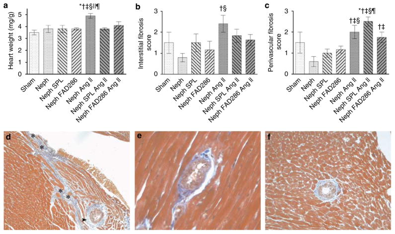
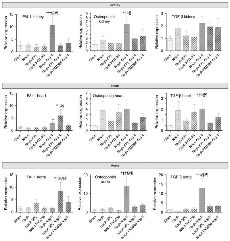
In the setting of high salt intake, aldosterone stimulates fibrosis in the heart, great vessels, and kidney of rats. We used uninephrectomized rats treated with angiotensin II and placed on a high salt diet to exaggerate renal fibrosis. We then tested whether mineralocorticoid receptor blockade by spironolactone or aldosterone synthase inhibition by FAD286 have similar effects on end-organ damage and gene expression. Individually, both drugs prevented the hypertensive response to uninephrectomy and high salt intake but not when angiotensin II was administered. Following 4 weeks of treatment with FAD286, plasma aldosterone was reduced, whereas spironolactone increased aldosterone at 8 weeks of treatment. Angiotensin II and high salt treatment caused albuminuria, azotemia, renovascular hypertrophy, glomerular injury, increased plasminogen activator inhibitor-1 (PAI-1), and osteopontin mRNA expression, as well as tubulointerstitial fibrosis in the kidney. Both drugs prevented these renal effects and attenuated cardiac and aortic medial hypertrophy while reducing osteopontin and transforming growth factor-beta mRNA expression in the aorta. The two drugs also reduced cardiac interstitial fibrosis but had no effect on that of the perivascular region. Although spironolactone enhanced angiotensin II and salt-stimulated PAI-1 mRNA expression in aorta and heart, spironolactone and FAD286 prevented renal PAI-1 mRNA protein expression. Our study shows that mineralocorticoid receptor antagonism and aldosterone synthase inhibition similarly decrease hypertrophy and interstitial fibrosis of the kidney and heart caused by angiotensin II and high salt.
Figures


References
-
- Brilla CG, Matsubara LS, Weber KT. Anti-aldosterone treatment and the prevention of myocardial fibrosis in primary and secondary hyperaldosteronism. J Mol Cell Cardiol. 1993;25:563–575. - PubMed
-
- Benetos A, Lacolley P, Safar ME. Prevention of aortic fibrosis by spironolactone in spontaneously hypertensive rats. Arterioscler Thromb Vasc Biol. 1997;17:1152–1156. - PubMed
-
- Rocha R, Rudolph AE, Frierdich GE, et al. Aldosterone induces a vascular inflammatory phenotype in the rat heart. Am J Physiol Heart Circ Physiol. 2002;283:H1802–H1810. - PubMed
Publication types
MeSH terms
Substances
Grants and funding
LinkOut - more resources
Full Text Sources
Other Literature Sources
Medical
Research Materials
Miscellaneous
