. 2009 Jun;75(11):1166-1172.
doi: 10.1038/ki.2009.24. Epub 2009 Feb 18.

Reversal of mineral ion homeostasis and soft-tissue calcification of klotho knockout mice by deletion of vitamin D 1alpha-hydroxylase

Affiliations

Reversal of mineral ion homeostasis and soft-tissue calcification of klotho knockout mice by deletion of vitamin D 1alpha-hydroxylase

Mutsuko Ohnishi et al. Kidney Int. 2009 Jun.

Abstract

Changes in the expression of klotho, a beta-glucuronidase, contribute to the development of features that resemble those of premature aging, as well as chronic renal failure. Klotho knockout mice have increased expression of the sodium/phosphate cotransporter (NaPi2a) and 1alpha-hydroxylase in their kidneys, along with increased serum levels of phosphate and 1,25-dihydroxyvitamin D. These changes are associated with widespread soft-tissue calcifications, generalized tissue atrophy, and a shorter lifespan in the knockout mice. To determine the role of the increased vitamin D activities in klotho knockout animals, we generated klotho and 1alpha-hydroxylase double-knockout mice. These double mutants regained body weight and developed hypophosphatemia with a complete elimination of the soft-tissue and vascular calcifications that were routinely found in klotho knockout mice. The markedly increased serum fibroblast growth factor 23 and the abnormally low serum parathyroid hormone levels, typical of klotho knockout mice, were significantly reversed in the double-knockout animals. These in vivo studies suggest that vitamin D has a pathologic role in regulating abnormal mineral ion metabolism and soft-tissue anomalies of klotho-deficient mice.


Conflict of interest statement

DISCLOSURE All the authors declared no competing interests.

Figures

Figure 1
Figure 1. Analysis of vitamin D components
Renal expression of 1α-hydroxylase enzyme (1α(OH)ase) and serum levels of 1,25-dihydroxyvitamin D (1,25(OH)2D3) in wild-type (WT) control and klotho−/− mice. Compared with the control mice (n =6), expression of 1α(OH)ase, an enzyme that converts inactive vitamin D components to the active 1,25(OH)2D3, was found to be elevated in kidneys obtained from klotho−/− mice (n =10), as determined by real-time PCR (1±0.27 wild-type vs 20.53±3.9 klotho−/−). Note that, compared with control mice (n =8; 38.3±7.5 pmol/l), serum 1,25(OH)2D3 levels were also significantly elevated in klotho−/− mice (n =8; 150.25±19.4 pmol/l) (*P<0.001 vs wild-type).
Figure 2
Figure 2. Macroscopic phenotype of klotho−/−/1α(OH)ase−/− double mutants
Gross phenotype of wild-type, klotho−/−, klotho−/−/1α(OH)ase−/− (DKO), and 1α(OH)ase−/− mice at around 9 weeks of age (upper panel). Body weight curves (lower panel) for wild-type (WT; n =12), klotho−/− (n =19), DKO (n =8), and 1α(OH)ase−/− (n =30) mice show that DKO mice are smaller than wild-type mice but larger than klotho−/− mice. This suggests that inactivation of vitamin D function in klotho−/− mice aided in body weight gains in DKO mice (*P<0.05, klotho−/− mice vs DKO mice; **P<0.001, klotho−/− mice vs DKO mice; #P<0.01, wild-type mice vs klotho−/− mice; ##P<0.001, wild-type mice vs klotho−/− mice).
Figure 3
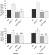
Figure 3. Biochemical measurement of serum phosphate and calcium in various genotypes
Note that the serum phosphate (upper panel) and calcium (lower panel) levels are higher in klotho−/− mice compared with wild-type (WT) mice. Compared with WT mice (n =7; 6.6±0.18), the serum phosphate level was significantly higher in klotho−/− mice (n =5; 11.1±0.39) at 3 weeks of age. Similar hyperphosphatemia was also observed in klotho−/− mice (n =12; 12.5±0.73) at 6 weeks of age, compared with WT mice (n =11; 7.2±0.35) of same age. In contrast to klotho−/− mice, the serum phosphate level was significantly reduced in klotho−/−/1α(OH)ase−/− double-knockout (DKO) mice, both at 3 weeks (n =6; 4.9±0.28) and 6 weeks (n =6; 4.6±0.27) of age. Similar reduction of serum phosphate level was also observed in 1α(OH)ase−/− mice at 3 weeks (n =5; 5.3±0.67) and 6 weeks (n =8; 5.3±0.26) of age. As for serum calcium (lower panel), compared with the WT mice (n =7; 7.4±0.1), the serum calcium level was significantly higher in klotho−/− mice (n =6; 9±0.42) at 3 weeks of age. Similar higher serum level of calcium was also observed in klotho−/− mice (n =6; 10.5±0.36) at 6 weeks of age, compared with the WT mice (n =9; 8.4±0.42) of same age. In contrast to the klotho−/− mice, the serum calcium level was significantly reduced in DKO mice, both at 3 weeks (n =6; 6.4±0.32) and 6 weeks (n = 6; 5.6±0.3) of age. Slightly reduced serum calcium level was also observed in 1α(OH)ase−/− mice at 3 weeks (n =6; 6.7±0.56) and 6 weeks (n =6; 6.1±0.57) of age (*P<0.001 vs WT; **P<0.001 vs klotho−/−; ***P<0.001 vs klotho−/−; #P<0.01 vs WT; ##P<0.001 vs klotho−/−; ###P<0.001 vs klotho−/−).
Figure 4
Figure 4. von Kossa staining of kidney tissues
Renal sections prepared from wild-type (WT), klotho−/−, klotho−/−/1α(OH)ase−/− double-knockout (DKO), and 1α(OH)ase−/− mice showing extensive calcifications in the kidneys of klotho−/− mice. Inactivation of vitamin D from klotho−/− mice completely eliminated calcification from DKO mice (magnification ×20). The macroscopic features of corresponding kidneys are shown in the upper panel.
Figure 5
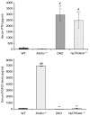
Figure 5. Biochemical measurements of serum PTH and Fgf23 in various genotypes
Note that compared with wild-type mice (n =4; 102.5±69), serum parathyroid hormone (PTH) levels are markedly reduced in klotho−/− mice (n =8; 46.8±16) but are significantly higher in both klotho−/−/1α(OH)ase−/− double-knockout (DKO) mice (n =4; 2945±560) and 1α(OH)ase−/− mice (n = 4; 2443±610). The average serum levels of fibroblast growth factor 23 (FGF23) are higher in klotho−/− mice (n =5; 6857 pg/ml), as compared with the wild-type mice (n =5; 134 pg/ml). In contrast to the klotho−/− mice, serum FGF23 levels are significantly reduced in DKO mice, (n =5; 6.7 pg/ml). The average serum levels of FGF23 in 1α(OH)ase−/− mice (n =5; 120 pg/ml) are also lower than in the klotho−/− mice (#P<0.01 vs wild-type; ##P<0.001 vs wild-type; **P<0.001 vs klotho−/−).
Figure 6
Figure 6. Histological analysis of lung tissues
Hematoxylin and eosin-stained sections of the lung tissues of 6-week-old wild-type (WT), klotho−/−, klotho−/−/1α(OH)ase−/− double-knockout (DKO), and 1α(OH)ase−/− mice. Note that, compared with wild-type mice, there is marked expansion of alveolar spaces (emphysema) in klotho−/− mice. Such pulmonary emphysematous changes are seen neither in the DKO mice nor in the 1α(OH)ase−/− mice (lung, magnification ×10).
Figure 7
Figure 7. Histological analysis of intestine
Compared with wild-type (WT) mice, there is marked atrophy of the intestinal wall (arrows) in klotho−/− mice. Such atrophic changes of the intestine are seen neither in the klotho−/−/1α(OH)ase−/− double-knockout (DKO) mice nor in the 1α(OH)ase−/− single-knockout mice (intestine, magnification ×10).

Comment in

References

    1. Lanske B, Razzaque MS. Vitamin D and aging: old concepts and new insights. J Nutr Biochem. 2007;18:771–777. - PMC - PubMed
    1. Kuro-o M, Matsumura Y, Aizawa H. Mutation of the mouse klotho gene leads to a syndrome resembling ageing. Nature. 1997;390:45–51. - PubMed
    1. Nakatani T, Bara S, Ohnishi M, et al. In vivo genetic evidence for klotho-dependent, fibroblast growth factor (FGF23)-mediated regulation of systemic phosphate homeostasis. FASEB J. 2009;23:433–441. - PMC - PubMed
    1. Tsujikawa H, Kurotaki Y, Fujimori T, et al. Klotho, a gene related to a syndrome resembling human premature aging, functions in a negative regulatory circuit of vitamin D endocrine system. Mol Endocrinol. 2003;17:2393–2403. - PubMed
    1. Memon F, El-Abbadi M, Nakatani T, et al. Does Fgf23-klotho activity influence vascular and soft tissue calcification through regulating mineral ion metabolism? Kidney Int. 2008;74:566–570. - PMC - PubMed

Publication types