. 2008 Dec 15;2(2):155-67.
doi: 10.3389/neuro.01.026.2008. eCollection 2008 Dec.

Many specialists for suppressing cortical excitation

Affiliations

Many specialists for suppressing cortical excitation

Andreas Burkhalter. Front Neurosci. .

Abstract

Cortical computations are critically dependent on GABA-releasing neurons for dynamically balancing excitation with inhibition that is proportional to the overall level of activity. Although it is widely accepted that there are multiple types of interneurons, defining their identities based on qualitative descriptions of morphological, molecular and physiological features has failed to produce a universally accepted 'parts list', which is needed to understand the roles that interneurons play in cortical processing. A list of features has been published by the Petilla Interneurons Nomenclature Group, which represents an important step toward an unbiased classification of interneurons. To this end some essential features have recently been studied quantitatively and their association was examined using multidimensional cluster analyses. These studies revealed at least 3 distinct electrophysiological, 6 morphological and 15 molecular phenotypes. This is a conservative estimate of the number of interneuron types, which almost certainly will be revised as more quantitative studies will be performed and similarities will be defined objectively. It is clear that interneurons are organized with physiological attributes representing the most general, molecular characteristics the most detailed and morphological features occupying the middle ground. By themselves, none of these features are sufficient to define classes of interneurons. The challenge will be to determine which features belong together and how cell type-specific feature combinations are genetically specified.

Keywords: GABAergic neurons; cerebral cortex; inhibition; interneurons.


Figures

Figure 1
Figure 1
Six distinct morphological types of interneurons. Diagrams showing different morphological types of interneurons (red), which were identified by cluster analyses of the subcellular distribution of axonal projections onto somata and dendrites of postsynaptic pyramidal neurons (gray). Chandelier cells (ChC), small basket cells (SBC), large basket cells (LBC), neurogliaform cells (NGFC), double bouquet cells (DBC) and Martinotti cells (MC). For examples of non-diagrammatic reconstructions of interneurons from rat somatosensory cortex and monkey prefrontal cortex see Krimer et al. (2005), Huang et al. (2007) and Ascoli et al. (2008).
Figure 2
Figure 2
Associations of electrophysiological and morphological properties of interneurons based on cluster analyses. Left side: Fast spiking (FS) properties (i.e., narrow action potentials, non-accommodating) are strongly linked (indicated by thick lines) to chandelier cells (ChC) and basket cells (BC). Relatively few (indicated by thin lines) interneurons show regular spiking (RS) properties (i.e., broad action potentials, accommodating), which are typically found in pyramidal cells. Intermediate spiking (IS) properties (i.e., narrow action potentials, accommodating) are strongly associated with interneurons that have vertically oriented axons (e.g., double bouquet cells (DBC) and Martinotti cells (MC)). Neurogliaform cells (NGC) never express FS properties. Right side: BCs receive high amplitude spontaneous excitatory postsynaptic potentials (sEPSP) and generate fast decaying spontaneous inhibitory postsynaptic potentials (sIPSPs). Putative DBCs and/or NGCs receive medium amplitude sEPSPs and more slowly decaying sIPSPs. Notice, because the cells have only been identified by the absence of BC and MC morphologies, the associations are indicated by dashed lines. MCs receive low amplitude sEPSPs and generate slowly decaying sIPSPs.
Figure 3
Figure 3
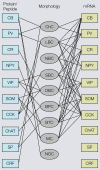
Associations of molecular (protein, mRNA) and morphological properties of interneurons. Left side: Immunocytochemical expression of commonly used markers [i.e., calbindin (CB), parvalbumin (PV), calretinin (CR), neuropeptide Y (NPY), vasointestinal polypeptide (VIP), somatostatin (SOM), cholecystokinin (CCK), choline acetyltransferase (ChAT), substance P (SP) corticotropin releasing factor (CRF)] in different morphological types of interneurons [i.e., chandelier cells (ChC), large basket cells (LBC), nest basket cells (NBC), small basket cells (SBC), bipolar cells (BPC), bitufted cells (BTC), Martinotti cells (MC), neurogliaform cells (NGC)]. Right side: mRNAs expression in different types of interneurons. Notice that most substances are coexpressed in multiple cell types and that the overlap in mRNA expression is much more extensive than immunostaining, suggesting that antibody staining is a more discriminating classification tool. Thus, genotypic and phenotypic molecular classification schemes are not interchangeable.
Figure 4
Figure 4
Inhibitory networks involving fast spiking parvalbumin (PV)-expressing neurons in thalamocortical, interlaminar and interareal cortical circuits. Feedforward excitatory thalamocortical inputs to spiny neurons (▴) and fast spiking interneurons (•) in layers 2–4 (a). Inputs to interneurons are stronger (large arrowheads) than inputs to spiny cells. PV neurons provide strong (large rectangular endings) feedforward inhibition (b) to spiny cells. Feedback inhibition (c) results from PV neurons that are excited by the same spiny neurons that they inhibit. These reciprocally connected spiny neuron/PV neuron pairs share common inputs (e.g., cells in layer 4 from thalamus or cells in layer 2/3 from layer 4) creating recurrent excitatory (d) and inhibitory subnetworks (contained within blue shaded boxes). ‘Lateral’ inhibition (e) of these subnetworks results from PV neurons that are driven by excitatory feedback connections (f) from outside the subnetworks (e.g., by layer 5 to layer 2/3 connections or feedback from higher cortical areas). Notice that ‘lateral’ inhibition is weaker (small rectangular endings) than feedforward and feedback inhibition and impinges on multiple subnetworks.

References

    1. Agmon A., Connors B. W. (1992). Correlation between intrinsic firing patterns and thalamocortical synaptic responses of neurons in mouse barrel cortex. J. Neurosci. 12, 319–329 - PMC - PubMed
    1. Ahmed B., Anderson J. C., Douglas R. J., Martin K. A. C., Nelson J. C. (1994). Polyneuronal innervation of spiny stellate neurons in cat visual cortex. J. Comp. Neurol. 341, 39–4910.1002/cne.903410105 - DOI - PubMed
    1. Angulo M. C., Rossier J., Staiger J. F., Audinat E. (1999). Postsynaptic glutamate receptors and integrative properties of fast-spiking interneurons in the rat neocortex. J. Neurophysiol. 82, 1295–1302 - PubMed
    1. Ascoli G. A., Alonso-Nanclares L., Anderson S. A., Barrinuevo G., Benavides-Piccone R., Burkhalter A., et al. (2008). Petilla terminology: nomenclature of features of GABAergic interneurons of the cerebral cortex. Nat. Rev. Neurosci. 9, 1–1210.1038/nrn2402 - DOI - PMC - PubMed
    1. Baraban S. C., Tallent M. K. (2004). Interneurons diversity series: Interneuronal neuropeptides – endogenous regulators of neuronal excitability. Trends Neurosci. 27, 135–14210.1016/j.tins.2004.01.008 - DOI - PubMed

LinkOut - more resources