Review
. 2009 Mar;276(6):1506-16.
doi: 10.1111/j.1742-4658.2009.06909.x. Epub 2009 Feb 18.

Ribonuclease H: properties, substrate specificity and roles in retroviral reverse transcription

Affiliations
Review

Ribonuclease H: properties, substrate specificity and roles in retroviral reverse transcription

James J Champoux et al. FEBS J. 2009 Mar.

Abstract

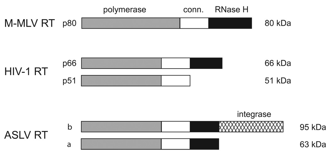
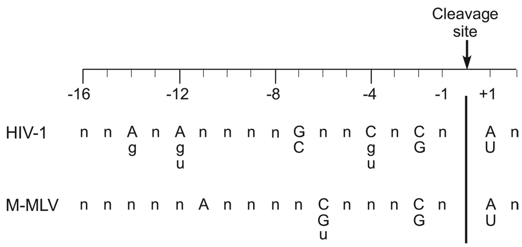
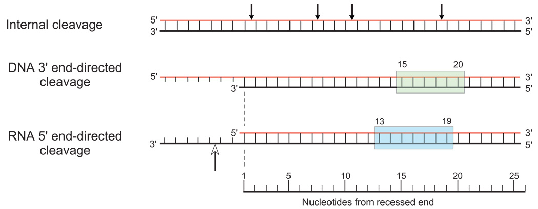
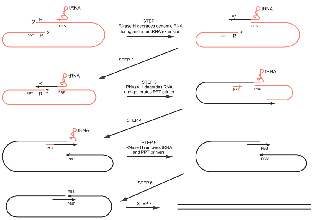
Retroviral reverse transcriptases possess both a DNA polymerase and an RNase H activity. The linkage with the DNA polymerase activity endows the retroviral RNases H with unique properties not found in the cellular counterparts. In addition to the typical endonuclease activity on a DNA/RNA hybrid, cleavage by the retroviral enzymes is also directed by both DNA 3' recessed and RNA 5' recessed ends, and by certain nucleotide sequence preferences in the vicinity of the cleavage site. This spectrum of specificities enables retroviral RNases H to carry out a series of cleavage reactions during reverse transcription that degrade the viral RNA genome after minus-strand synthesis, precisely generate the primer for the initiation of plus strands, facilitate the initiation of plus-strand synthesis and remove both plus- and minus-strand primers after they have been extended.


Figures

Fig. 1
Fig. 1
Subunit and domain structure of retroviral reverse transcriptases. Reverse transcriptase from M-MLV is a monomer, whereas the HIV-1 and ASLV reverse transcriptases are both heterodimeric. The subunit designations and their sizes (kDa) are indicated along the left and right sides of the figure, respectively. The approximate sizes of the polymerase, connection (conn.) and RNase H domains are shown in gray, white, and black, respectively. The larger β subunit of the ASLV reverse transcriptase also contains the integrase domain depicted by crosshatching.
Fig. 2
Fig. 2
Ribbon diagram of the co-crystal structure of HIV-1 reverse transcriptase with a bound RNA template and DNA primer (pdb entry 1HYS) [19]. The polymerase (p66 residues 1–318), connection (p66 residues 319–437) and RNase H (p66 residues 438–553) domains are drawn in red, green, and blue, respectively with the p51 subunit shown in gray. The RNase H active site is indicated with the four key acidic residues drawn in yellow ball and stick. The primer terminus of the DNA primer strand (purple) is indicated with the RNA template strand shown in yellow. The drawing was created using Swiss-Pdb Viewer software (v3.7) (GlaxoSmithKline).
Fig. 3
Fig. 3
Sequence preferences for internal cleavage by retroviral RNases H. For the purposes of site alignment, RNase H cleavage is designated as occurring between nucleotides −1 and +1. The preferred nucleotides at positions −14, −12, −7, −4, −2 and +1 are shown for HIV-1 RNase H and at positions −11, −6, −2 and +1 for M-MLV RNase H. The strongest preferences are indicated in upper case letters with the weaker preferences in lower case letters.
Fig. 4
Fig. 4
Three cleavage modes for retroviral RNases H. DNA/RNA hybrids are drawn with RNA strands in red and DNA strands in black. In the internal cleavage mode, the arrows mark the sites of cleavage along the length of the hybrid where nucleotide sequence alone determines the cleavage site. The cleavage window for the DNA 3' end-directed cleavage mode (15–20 nucleotides from the recessed DNA end) is highlighted in green. The corresponding cleavage window for RNA 5' end-directed cleavage (13–19 nucleotides from end) is highlighted in blue. The open-headed arrow in the RNA 5' end-directed cleavage mode indicates the position of the DNA phosphate that appears to be bound near the active site pocket in the polymerase domain normally occupied by the 3' DNA primer terminus during DNA polymerization.
Fig. 5
Fig. 5
Roles of RNase H in reverse transcription. The retroviral genome and the associated cell-derived tRNA bound to the primer binding site (PBS) are shown in red with the DNA strands produced during reverse transcription shown in black. A repeated sequence denoted R is located at both ends of the retroviral genome. The sequences complementary to PBS and R are denoted PBS' and R', respectively. The polypurine tract (PPT) serves as the primer for plus-strand synthesis. The steps at which RNase H plays a role are highlighted. See the text for a detailed explanation.

References

    1. Baltimore D. RNA-dependent DNA polymerase in virions of RNA tumour viruses. Nature. 1970;226:1209–1211. - PubMed
    1. Temin HM, Mizutani S. RNA-dependent DNA polymerase in virions of Rous sarcoma virus. Nature. 1970;226:1211–1213. - PubMed
    1. Molling K, Bolognesi DP, Bauer H, Busen W, Plassmann HW, Hausen P. Association of viral reverse transcriptase with an enzyme degrading the RNA moiety of RNA-DNA hybrids. Nat New Biol. 1971;234:240–243. - PubMed
    1. Champoux JJ. In: Reverse Transcriptase. Skalka AM, Goff SP, editors. Cold Spring Harbor, NY: Cold Spring Harbor Press; 1993. pp. 103–118.
    1. Telesnitsky A, Goff SP. In: Reverse Transcriptase. Skalka AM, Goff SP, editors. Plainview, New York: Cold Spring Harbor Laboratory Press; 1993. pp. 49–83.

Publication types