Review
. 2009 Mar;276(6):1494-505.
doi: 10.1111/j.1742-4658.2009.06908.x. Epub 2008 Feb 18.

Ribonuclease H: the enzymes in eukaryotes

Affiliations
Review

Ribonuclease H: the enzymes in eukaryotes

Susana M Cerritelli et al. FEBS J. 2009 Mar.

Abstract

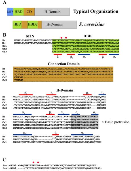
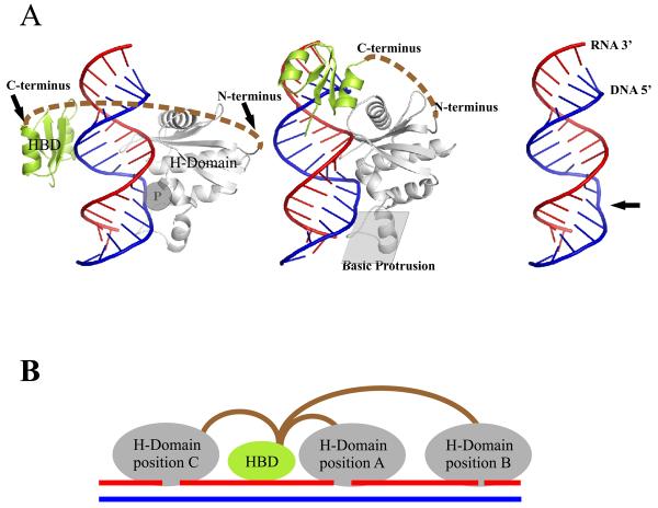
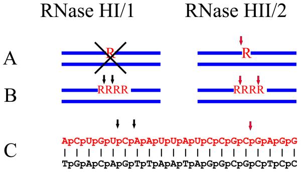
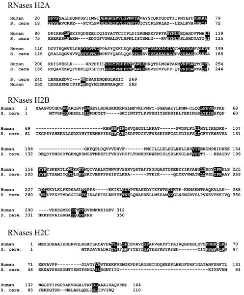
Ribonucleases H are enzymes that cleave the RNA of RNA/DNA hybrids that form during replication and repair and which could lead to DNA instability if they were not processed. There are two main types of RNase H, and at least one of them is present in most organisms. Eukaryotic RNases H are larger and more complex than their prokaryotic counterparts. Eukaryotic RNase H1 has acquired a hybrid binding domain that confers processivity and affinity for the substrate, whereas eukaryotic RNase H2 is composed of three different proteins: the catalytic subunit (2A), similar to the monomeric prokaryotic RNase HII, and two other subunits (2B and 2C) that have no prokaryotic counterparts and as yet unknown functions, but that are necessary for catalysis. In this minireview, we discuss some of the most recent findings on eukaryotic RNases H1 and H2, focusing on the structural data on complexes between human RNase H1 and RNA/DNA hybrids that had provided great detail of how the hybrid binding- and RNase H-domains recognize and cleave the RNA strand of the hybrid substrates. We also describe the progress made in understanding the in vivo function of eukaryotic RNases H. Although prokayotes and some single-cell eukaryotes do not require RNases H for viability, in higher eukaryotes RNases H are essential. Rnaseh1 null mice arrest development around E8.5 because RNase H1 is necessary during embryogenesis for mitochondrial DNA replication. Mutations in any of the three subunits of human RNase H2 cause Aicardi-Goutières syndrome, a human neurological disorder with devastating consequences.


Figures

Figure 1
Figure 1
RNase H1 organization in eukaryotes. (A) Two types of RNases H1 in eukaryotes are shown. The top “typical organization” is found in most RNases H1 from higher eukaryotes and consists of an MTS (Mitochondrial Targeting Sequence), HBD (Hybrid Binding Domain), CD (Connection Domain) and H-Domain (RNase H Domain). Not all “typical” RNases H1 have an MTS. Some, mainly fungal, RNases H1 have two HBDs as shown for S. cerevisiae. (B) Alignments of human (Hs), mouse (Mo), and three RNase H1 type proteins encoded in C. albicans (Ca1-3). The red bars represent the β-strands of human RNase H1, and the blue bars are the α-helices. The basic protrusion is shaded in grey as noted in the H-Domain alignment. (C) Alignment of the HBDs of S. cerevisiae RNase H1. Red circles in C and B are the partially conserved HBD amino acids discussed in the text.
Figure 2
Figure 2
Structural model. Interplay of HBD and H-Domain is orchestrated by the connection domain. (A) Co-crystal structure of human RNase H1 HBD and RNase H domain with RNA/DNA. Left and middle panels show HBD modeled either on the opposite side of RNase H domain (left) or immediately upstream (middle). The C-terminus of the HBD is connected to the N-terminus of the RNase H domain as indicated by dashed brown lines. The phosphate binding pocket is indicated as a “P” inside a grey circle. The middle panel also highlights the position of the basic protrusion. The right panel shows the RNA/DNA with the protein removed revealing the bend in the DNA marked by an arrow. (B) Processivity of RNase H1. In the model of processivity of RNase H1, the HBD anchors the enzyme to a hybrid permitting the RNase H domain to engage with several sites on a single RNA/DNA leading to multiple cleavages from a single binding event. In position A of the H-Domain, hydrolysis will occur near the HBD while in position B cleavage will be more distal. To achieve binding and cleavage at position C, the connection domain needs to be flexible to change the relative orientation of the HBD and H-Domains although the orientation with respect to the hybrid must remain the same.
Figure 3
Figure 3
RNase HI/1 and RNase HII/2 have distinct cleavage patterns on three different substrates. Three types of substrates are shown. (A) A single ribonucleotide in a duplex DNA that is cleaved by RNases HII/2 but not RNases HI/1 as noted by X. (B) Four consecutive ribonucleotides residues and (C) an RNA/DNA hybrid that is cleaved differently by the two classes of enzymes. Black arrows denote sites of cleavage by RNase HI/1 and red arrows represent hydrolysis by RNase HII/2.
Figure 4
Figure 4
(A) Comparison of amino acid sequences of human and S. cerevisiae RNase H2 subunits. Alignments are based on the consensus alignment of RNase H2 subunits from Crow et al. Reverse letters highlight homologies between the subunits. For RNases H2B and RNases H2C, a more generous highlighting is used and even then it is clear there is very little conservation between the human and yeast B and C subunits. (B) Schematic representation of the three components of human RNase H2. On top of each subunit are shown AGS-related mutations, and on the bottom the amino acids involved in catalysis, in RNASEH2A, and the PIP (PCNA Interacting Peptide) site of RNASEH2B.
Figure 4
Figure 4
(A) Comparison of amino acid sequences of human and S. cerevisiae RNase H2 subunits. Alignments are based on the consensus alignment of RNase H2 subunits from Crow et al. Reverse letters highlight homologies between the subunits. For RNases H2B and RNases H2C, a more generous highlighting is used and even then it is clear there is very little conservation between the human and yeast B and C subunits. (B) Schematic representation of the three components of human RNase H2. On top of each subunit are shown AGS-related mutations, and on the bottom the amino acids involved in catalysis, in RNASEH2A, and the PIP (PCNA Interacting Peptide) site of RNASEH2B.

References

    1. Stein H, Hausen P. Enzyme from Calf Thymus Degrading the RNA Moiety of DNA-RNA Hybrids: Effect on DNA-Dependent RNA Polymerase. Science. 1969;166:393–395. - PubMed
    1. Büsen W, Frank P. Bovine RNases H. In: Crouch RJ, Toulmé JJ, editors. Ribonucleases H. INSERM; Paris: 1998. pp. 113–146.
    1. Itaya M. Isolation and characterization of a second RNase H (RNase HII) of Escherichia coli K-12 encoded by the rnhB gene. Proc. Natl. Acad. Sci.U.S.A. 1990;87:8587–8591. - PMC - PubMed
    1. Crouch RJ, Arudchandran A, Cerritelli SM. RNase H1 of Saccharomyces cerevisiae: methods and nomenclature. Methods Enzymol. 2001;341:395–413. 395-413. - PubMed
    1. Ohtani N, Haruki M, Morikawa M, Crouch RJ, Itaya M, Kanaya S. Identification of the genes encoding Mn2+-dependent RNase I-III and Mg2+-dependent RNase HIII from Bacillus subtilis: Classification of RNases H into three families. Biochemistry. 1999;38:605–618. - PubMed

Publication types