

Expression of Bmi-1 is a prognostic marker in bladder cancer
- PMID: 19228380
- PMCID: PMC2652492
- DOI: 10.1186/1471-2407-9-61
Expression of Bmi-1 is a prognostic marker in bladder cancer
Abstract
Background: The molecular mechanisms of the development and progression of bladder cancer are poorly understood. The objective of this study was to analyze the expression of Bmi-1 protein and its clinical significance in human bladder cancer.
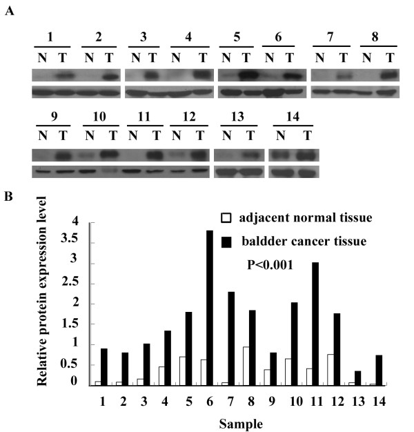
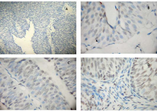
Methods: We examined the expression of Bmi-1 mRNA and Bmi-1 protein by RT-PCR and Western blot, respectively in 14 paired bladder cancers and the adjacent normal tissues. The expression of Bmi-1 protein in 137 specimens of bladder cancer and 30 specimens of adjacent normal bladder tissue was determined by immunohistochemistry. Statistical analyses were applied to test the relationship between expression of Bmi-1, and clinicopathologic features and prognosis.
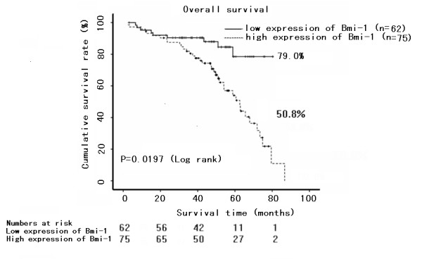
Results: Expression of Bmi-1 mRNA and protein was higher in bladder cancers than in the adjacent normal tissues in 14 paired samples (P < 0.01). By immunohistochemical examination, five of 30 adjacent normal bladder specimens (16.7%) versus 75 of 137 bladder cancers (54.3%) showed Bmi-1 protein expression (P < 0.05). Bmi-1 protein expression was intense in 20.6%, 54.3%, and 78.8% of tumors of histopathological stages G1, G2, and G3, respectively (P < 0.05). Expression of Bmi-1 protein was greater in invasive bladder cancers than in superficial bladder cancers (81.5% versus 32.5%, P < 0.05). In invasive bladder cancers, the expression of Bmi-1 protein in progression-free cancers was similar to that of cancers that have progressed (80.0% versus 82.4%, P > 0.5). In superficial bladder cancers, the expression of Bmi-1 protein in recurrent cases was higher than in recurrence-free cases (62.5% versus 13.7%, P < 0.05). Bmi-1 expression was positively correlated with tumor classification and TNM stage (P < 0.05), but not with tumor number (P > 0.05). Five-year survival in the group with higher Bmi-1 expression was 50.8%, while it was 78.5% in the group with lower Bmi-1 expression (P < 0.05). Patients with higher Bmi-1 expression had shorter survival time, whereas patients with lower Bmi-1 expression had longer survival time (P < 0.05).
Conclusion: Expression of Bmi-1 was greater in bladder cancers than in the adjacent normal tissues. The examination of Bmi-1 protein expression is potentially valuable in prognostic evaluation of bladder cancer.
Figures

References
-
- Laufer M, Ramalingam S, Schoenberg MP, Haisfield-Wolf ME, Zuhowski EG, Trueheart IN, Eisenberger MA, Nativ O, Egorin MJ. Intravesical Gemcitabine therapy for superficial transitional cell carcinoma of the bladder phase I and pharmacokinetic study. J Clin Oncol. 2003;21:697–703. doi: 10.1200/JCO.2003.09.028. - DOI - PubMed
Publication types
MeSH terms
Substances
LinkOut - more resources
Full Text Sources
Medical
