

Abnormal resting-state cortical coupling in chronic tinnitus
- PMID: 19228390
- PMCID: PMC2649130
- DOI: 10.1186/1471-2202-10-11
Abnormal resting-state cortical coupling in chronic tinnitus
Abstract
Background: Subjective tinnitus is characterized by an auditory phantom perception in the absence of any physical sound source. Consequently, in a quiet environment, tinnitus patients differ from control participants because they constantly perceive a sound whereas controls do not. We hypothesized that this difference is expressed by differential activation of distributed cortical networks.
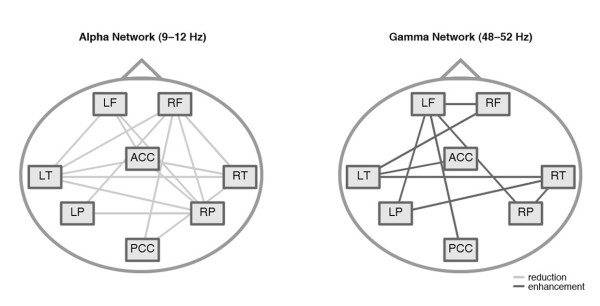
Results: The analysis was based on a sample of 41 participants: 21 patients with chronic tinnitus and 20 healthy control participants. To investigate the architecture of these networks, we used phase locking analysis in the 1-90 Hz frequency range of a minute of resting-state MEG recording. We found: 1) For tinnitus patients: A significant decrease of inter-areal coupling in the alpha (9-12 Hz) band and an increase of inter-areal coupling in the 48-54 Hz gamma frequency range relative to the control group. 2) For both groups: an inverse relationship (r = -.71) of the alpha and gamma network coupling. 3) A discrimination of 83% between the patient and the control group based on the alpha and gamma networks. 4) An effect of manifestation on the distribution of the gamma network: In patients with a tinnitus history of less than 4 years, the left temporal cortex was predominant in the gamma network whereas in patients with tinnitus duration of more than 4 years, the gamma network was more widely distributed including more frontal and parietal regions.
Conclusion: In the here presented data set we found strong support for an alteration of long-range coupling in tinnitus. Long-range coupling in the alpha frequency band was decreased for tinnitus patients while long-range gamma coupling was increased. These changes discriminate well between tinnitus and control participants. We propose a tinnitus model that integrates this finding in the current knowledge about tinnitus. Furthermore we discuss the impact of this finding to tinnitus therapies using Transcranial Magnetic Stimulation (TMS).
Figures


References
-
- Meyerhoff WL, Cooper JC. Otolaryngology. 3. Philadelphia: Saunders; 1991. Tinnitus; pp. 1169–75.
-
- Phoon WH, Lee HS, Chia SE. Tinnitus in noise-exposed workers. Occup Med (Lond) 1993;43:35–38. - PubMed
-
- Eggermont JJ, Roberts LE. The neuroscience of tinnitus. Trends Neurosci. 2004;27:676–682. - PubMed
-
- Silverstein H. Transmeatal labyrinthectomy with and without cochleovestibular neurectomy. Laryngoscope. 1976;86:1777–1791. - PubMed
Publication types
MeSH terms
LinkOut - more resources
Full Text Sources
Other Literature Sources
Medical
