. 2009 Feb 19;28(1):23.
doi: 10.1186/1756-9966-28-23.

A pilot study on acute inflammation and cancer: a new balance between IFN-gamma and TGF-beta in melanoma

Affiliations

A pilot study on acute inflammation and cancer: a new balance between IFN-gamma and TGF-beta in melanoma

Yue-mei Ma et al. J Exp Clin Cancer Res. .

Abstract

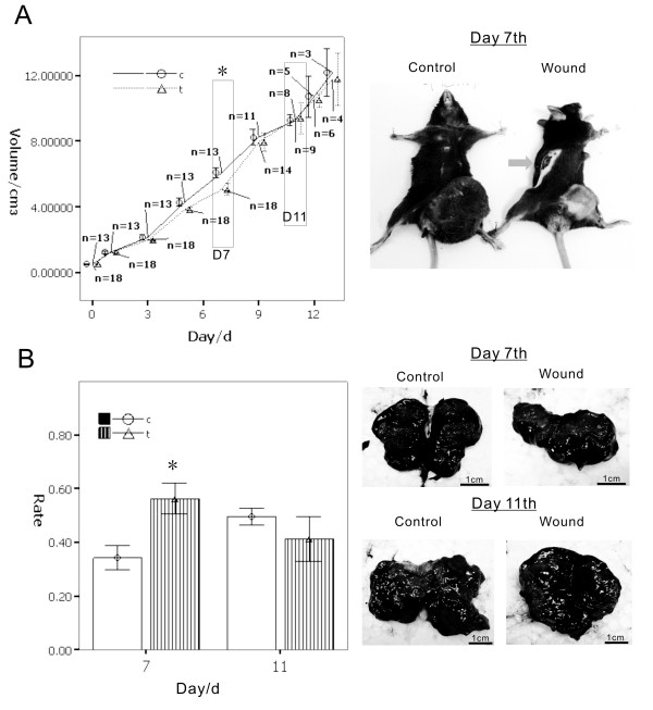
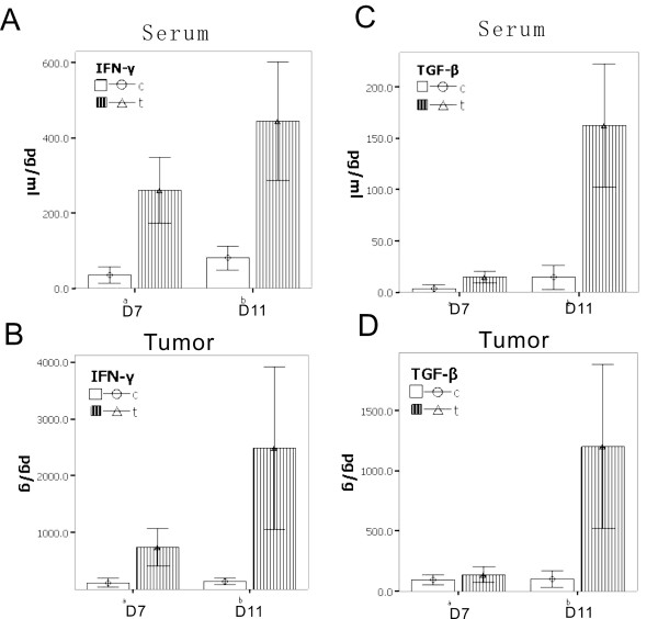
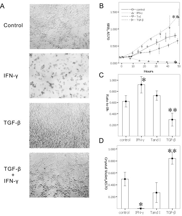
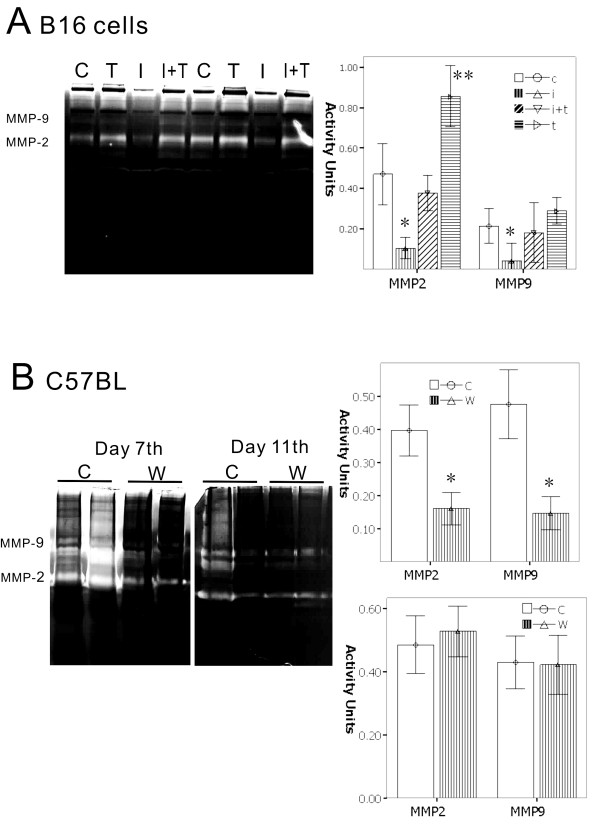
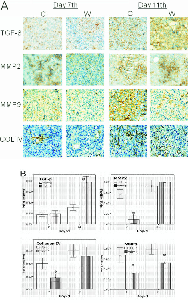
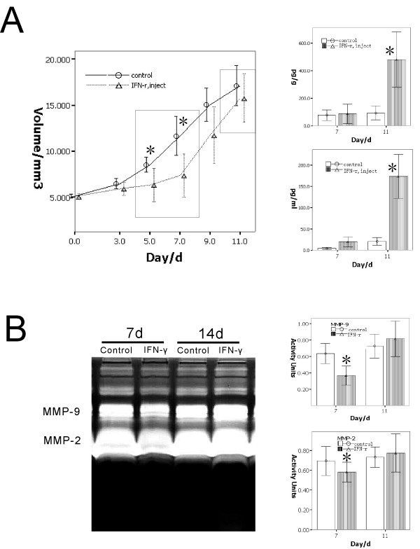
Recent data have redefined the concept of inflammation as a critical component of tumor progression. However, there has been little development on cases where inflammation on or near a wound and a tumor exist simultaneously. Therefore, this pilot study aims to observe the impact of a wound on a tumor, to build a new mouse tumor model with a manufactured surgical wound representing acute inflammation, and to evaluate the relationship between acute inflammation or wound healing and the process of tumor growth. We focus on the two phases that are present when acute inflammation influences tumor. In the early phase, inhibitory effects are present. The process that produces these effects is the functional reaction of IFN-gamma secretions from a wound inflammation. In the latter phase, the inhibited tumor is made resistant to IFN-gamma through the release of TGF-beta to balance the inflammatory factor effect on the tumor cells. A pair of cytokines IFN-gamma/TGF-beta established a new balance to protect the tumor from the interference effect of the inflammation. The tumor was made resistant to IFN-gamma through the release of TGF-beta to balance the inflammatory effect on the tumor cells. This balance mechanism that occurred in the tumor cells increased proliferation and invasion. In vitro and in vivo experiments have confirmed a new view of clinical surgery that will provide more detailed information on the evaluation of tumors after surgery. This study also provides a better understanding of the relationship between tumor and inflammation, as well as tumor cell attacks on inflammatory factors.


Figures

Figure 1
Figure 1
A wound model was built in C57BL/B16 tumor-bearing mouse to determine the influence on melanoma by inflammation. When the tumor grew to 0.5 cm3, we created a wound beyond the tumor in the opposite site of the mouse's body. A.) The results show gradual reduction of the tumor volume when the wound was building; the tumor volume reached the minimum at day 7 (shown in black box, p < 0.01). After day 7, the tumor inhibitory effect of the wound (inflammation) weakened gradually. On about day 11 of the inflammatory group compared with the control group, tumor volume almost as same as the control group at day 13 (shown in black box, p > 0.05). B.) The cross-section of the tumor showed that the tumor necrosis with hemorrhage occurred in different proportions of times and groups. On day 7, the group wound tumors were smaller than the control group, and the area with necrotic tissue is greater than the control group (p < 0.01). After 11 days, the tumor volume in the wound group was increased, but in the cross-section area of necrotic tissue rather than in the control group (p > 0.05). The necrotic percentage after day 11 showed the tumor through a mechanism to adapt the wounds caused by inflammation induced necrosis, promoted the emergence of proliferation.
Figure 2
Figure 2
To further observe and determine the inflammatory factors in the interaction between tumor and inflammation, results showed that: A.) the level of IFN-γ in the serum in the wound group continued a high level of expression (day 7 p < 0.01, day 11 p < 0.01); B.) in tumor tissue also detected high concentrations compared with the control group (day 7 p < 0.01, day 11 p < 0.01). Interestingly, at the 11th day, the tumor with the TGF-β increased, the result is that: C.) high levels of TGF-β can also be detected in the serum (day 7 p > 0.05, day 11 p < 0.01); D.) the same change in tumor (day 7 p > 0.05, day 11 p < 0.01). The TGF-β level before day 7 is not clear in terms of the present low of expression and secretion of tumor cells. That showed that at this time, the tumor does not have to go through the regulation of TGF-β to go against the ability of IFN-γ. When the IFN-γ-induces inhibition of tumor necrosis and persistence over a period, the role of TGF-β has been demonstrated, giving the tumor cells the ability to fight against the IFN-γ, so that the tumor cells could grow.
Figure 3
Figure 3
To investigate the cells deal with cytokines in vitro. A-B.) Morphology shows that TGF-β induced a rapid proliferation of cells, and cells presented a spindle-like shape. The IFN-γ group presented a reduction tendency on cell adhesion, the shape of cells present suspended or polygonal, lose normal B16 cells morphousorm. When given TGF-β and IFN-γ at the same time, cells returned to normal B16 cell shape, and cells also grew. C.) The results by wound healing assay showed that TGF-β confronting IFN-γ can promote migration. To treat cells only by IFN-γ inhibited cells migration. D.) Based on the Transwell invasion assay, IFN can inhibit cell migration, and inhibit cell invasion through Matrigel, and TGF-β has the opposite effect on cells to IFN-γ, and may have also an activity for inhibiting the IFN-γ activity, so that the cells migrate and invade.
Figure 4
Figure 4
To verify whether TGF-β and IFN-γ can enhance melanoma cells invasion by gelatin zymography assay analyzed in vitro and in vivo. A.) B16 cells treated by cytokines, show that IFN-γ can reduce the activity of MMP-2 and MMP-9, which are key modulators of tumor invasion. On the other hand, TGF-β can enhance the activity of both MMP-2 and MMP-9, giving TGF-β and IFN-γ. At the same time, TGF-β confronted IFN-γ to recover the activity of MMPs, and performed increasing activities on MMP-2 and MMP-9. B.) In vivo animal experiments also showed that there are significant features in day 7; the wound group had significantly lower activities of MMP-2 and MMP-9 compared with the control group from, 30% to 50%, respectively. By day 11, there was no significant difference in the activity of MMP-2 and MMP-9 between the wound groups and the control group. (*, p < 0.01)
Figure 5
Figure 5
Based on wound model described above, the tumor sample's immunohistochemistry analysis showed that, the TGF-β positive cells both in the wound group and the control group day 7 presented weak expression. On day 11, the wound group presented significantly strong expression of positive cells higher than the control group. The positive cells of MMP-2 and MMP-9 show the same tendency as the results in the zymography, but when the TGF-β up-regulated expression, the activity of the state of MMP-2 and MMP-9 were restored from inhibiting to the highest expression. COL IV is an important extracellular matrix, and the percentage of positive cells in the wound group found on day 7 had a lower expression compared with the control group. However, in day 11, reflected in the control group with similar results, which show that both MMPs and extracellular matrix plasticity and inflammation will continue to dampen demand in the early phase, and reach to the latter phase. This is because the cytokines such as TGF-β, play new roles on tumor cells to escape the shackles of inflammatory factors, access to the growth, and progression. A.) The positive cells are stained in brown. B.) The positive percent of cells, p < 0.01 marked by *.
Figure 6
Figure 6
Determination of the effect of IFN-γ injection on the tumor via tail-vein to validate the IFN-γ released from the wound model. A.) The tumor growth curves showing the double-phase in the IFN-γ injection group, the inhibition phase, and the inhibition missing phase. In the inhibition missing phase, the level of TGF-β increased significantly in the IFN-γ injection group as compared to that in the control (marked by *, p < 0.05). B.) The activity of MMP-2 and MMP-9 as detected by the gelatin zymography analysis showing the decrease in the inhibition phase of the IFN-γ injection group and the significant increase in the inhibition missing phase as compared to the control group (marked by *, p < 0.05).

Similar articles

Cited by

References

    1. Balkwill F, Mantovani A. Inflammation and cancer: back to Virchow? Lancet. 2001;357:539–545. - PubMed
    1. Coussens LM, Werb Z. Inflammation and cancer. Nature. 2002;420:860–867. - PMC - PubMed
    1. Aggarwal BB, Shishodia S, Sandur SK, Pandey MK, Sethi G. Inflammation and cancer: how hot is the link? Biochem Pharmacol. 2006;72:1605–1621. - PubMed
    1. Chettibi S, Ferguson MW. In: Inflammation: Basic Principles and Clinical Correlates. Gallin JI, Snyderman R, editor. Williams and Wilkinson. Lipincott. Philadelphia; 1999. pp. 865–881.
    1. Brigati C, Noonan DM, Albini A, Benelli R. Tumors and inflammatory infiltrates: friends or foes? Clin Exp Metastasis. 2002;19:247–258. - PubMed

Publication types

Substances