. 2009 Jun;80(6):1136-45.
doi: 10.1095/biolreprod.108.075226. Epub 2009 Feb 18.

Isolation and culture of epithelial progenitors and mesenchymal stem cells from human endometrium

Affiliations

Isolation and culture of epithelial progenitors and mesenchymal stem cells from human endometrium

Caroline E Gargett et al. Biol Reprod. 2009 Jun.

Abstract

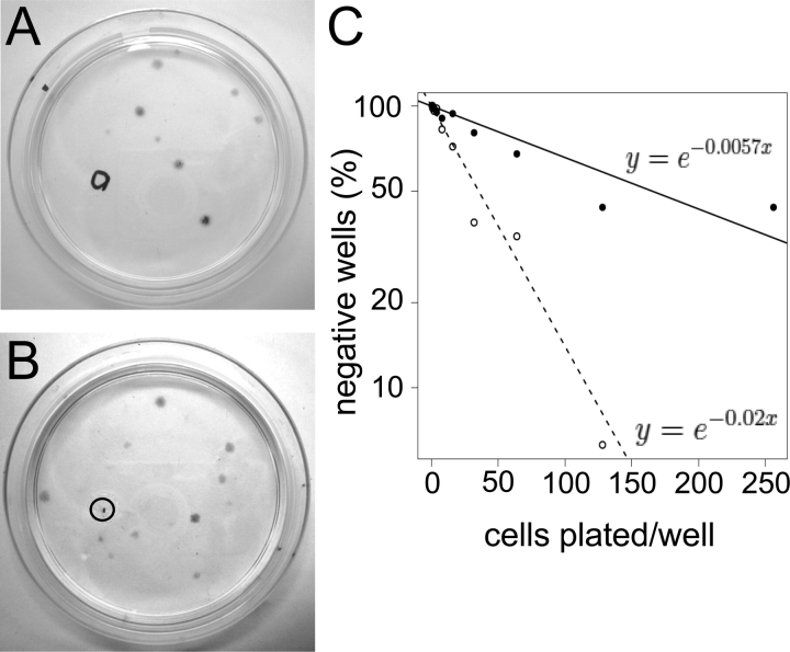
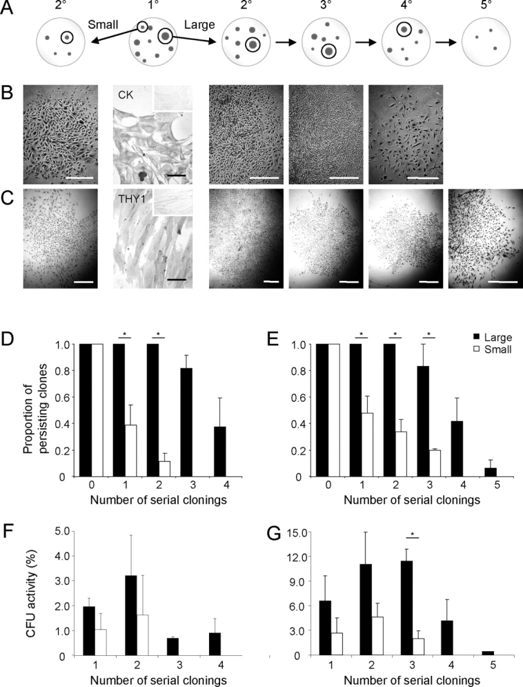
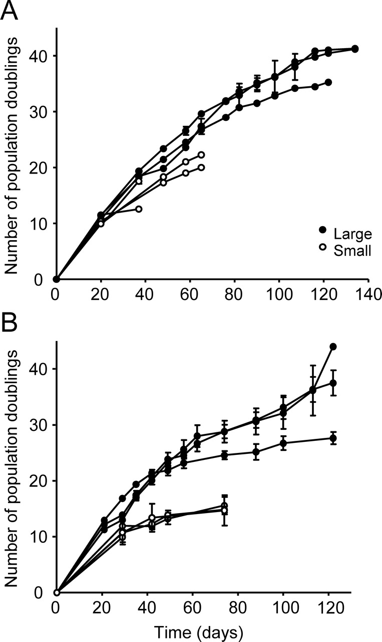
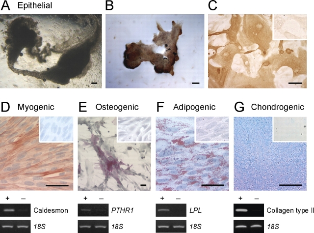
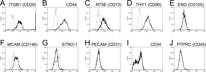
Human endometrium is a highly regenerative tissue undergoing more than 400 cycles of growth, differentiation, and shedding during a woman's reproductive years. Endometrial regeneration is likely mediated by adult stem/progenitor cells. This study investigated key stem cell properties of individual clonogenic epithelial and stromal cells obtained from human endometrium. Single-cell suspensions of endometrial epithelial or stromal cells were obtained from hysterectomy tissues from 15 women experiencing normal menstrual cycles, and were cultured at clonal density (10 cells/cm(2)) or limiting dilution. The adult stem cell properties-self-renewal, high proliferative potential, and differentiation of single epithelial and stromal cells-were assessed by harvesting individual colonies and undertaking serial clonal culture, serial passaging, and culture in differentiation-induction media, respectively. Lineage differentiation markers were examined by RT-PCR, immunocytochemistry, and flow cytometry. Rare single human endometrial EpCAM(+) epithelial cells and EpCAM(-) stromal cells demonstrated self-renewal by serially cloning >3 times and underwent >30 population doublings over 4 mo in culture. Clonally derived epithelial cells differentiated into cytokeratin(+) gland-like structures in three dimensional culture. Single stromal cells were multipotent, as their progeny differentiated into smooth muscle cells, adipocytes, chondrocytes, and osteoblasts. Stromal clones expressed mesenchymal stem cell (MSC) markers ITGB1 (CD29), CD44, NT5E (CD73), THY1 (CD90), ENG (CD105), PDGFRB (CD140B), MCAM (CD146) but not endothelial or hemopoietic markers PECAM1 (CD31), CD34, PTPRC (CD45). Adult human endometrium contains rare epithelial progenitors and MSCs, likely responsible for its immense regenerative capacity, which may also have critical roles in the development of endometriosis and endometrial cancer. Human endometrium may provide a readily available source of MSCs for cell-based therapies.


Figures

FIG. 1.
FIG. 1.
Human endometrial cell clones. Typical 6-cm cloning plates of freshly isolated single cell suspensions of epithelial (A) and stromal (B) cells seeded at 20 and 10 cells/cm2, respectively, showing well-separated individual colonies (CFU). Circles indicate removal of a clone for analysis. C) Limiting dilution analysis showing frequency of epithelial (black circles) and stromal (open circles) clones in endometrial cell suspensions by limiting dilution. Data are from four patient samples (nos. 11, 12, 14, and 15), with eight replicates/sample using Poisson distribution analysis. The frequency of stromal CFU was significantly greater than for epithelial (P < 0.0001).
FIG. 2.
FIG. 2.
Serial cloning analysis for measuring self-renewal of human endometrial large and small primary (1°) epithelial and stromal CFU. A) Schematic showing serial cloning strategy. The initial cloning plate (seeded at 10–20 cells/cm2), second panel with two cloning rings selecting the largest (>4000 cells), and a small to medium (<2000 cells) CFU. These clones were individually replated at 5–10 cells/cm2 and cultured for 14 days. Serial clonal passaging (2°–5°) is depicted as cloning plates containing representative clones, with selection of typical large clones for the subsequent round of cloning indicated within cloning rings, until CFU activity was exhausted (4°/5°). Typical endometrial epithelial (B) and stromal (C) colonies formed at each round of serial cloning. Endometrial epithelial colonies were cytokeratin+ (CK) and stromal colonies THY1 (CD90+) (second panels). Inserts are isotype controls. Rate of clonal extinction is shown for both large and small CFU for each round of serial cloning for epithelial (D) and stromal (E) CFU. Percentage of CFU in large and small epithelial (F) and stromal (G) CFU at each round of serial cloning. Results are means ± SEM (n = 3 patient samples [from nos. 7–9 for epithelial and nos. 1–4 for stromal); averages of three to five small and large CFU/cell type/patient sample). *Significant difference between large and small CFU (P < 0.05). Bars = 1 mm for clones (C); bars = 50 μm for immunostained images, including insets (B and C).
FIG. 3.
FIG. 3.
Proliferative potential of human endometrial large and small epithelial and stromal CFU. Growth curves from single primary large and small epithelial (A) and stromal (B) CFU from individual patient samples passaged at 2000 cells/cm2 illustrating differences in cell proliferation rates and total cell output. Each curve is the mean ± SEM of n = 3–5 clones derived from an individual patient sample (n = 3 patients, nos. 10, 12, and 13 for epithelial; n = 4 patients, nos. 1–4 for stromal).
FIG. 4.
FIG. 4.
Multilineage differentiation of single-cell-derived large human endometrial epithelial CFU and stromal CFU. AC) Clonally derived epithelial cells (large CFU) cultured in 50% Matrigel above an endometrial stromal cell feeder layer for 4 wk differentiated into epithelial gland-like structures (A) observed by phase contrast microscopy, and (B and C) immunoreactive for cytokeratin. Inset in C is the isotype control. DG) Clonally derived stromal cells (two clones) were cultured as monolayers or cell pellets (chondrogenic) for 4 wk in differentiation-induction media for mesenchymal lineages and assessed for lineage-specific markers by histochemistry, immunocytochemistry (upper panels), and RT-PCR (lower panels). D) Myogenic differentiation to smooth muscle cells, positive for αSMA and caldesmon. E) Osteogenic differentiation indicated by alkaline phosphatase reactivity and expression of parathyroid hormone receptor 1 (PTHR1). F) Adipogenic differentiation visualized by Oil Red O staining of lipid droplets and expression of lipoprotein lipase (LPL). G) Chondrogenic differentiation shown in a paraffin section of a micromass cell pellet stained with Alcian blue, and collagen type II expression. Cells cultured in control culture media for 4 wk and stained for lineage markers are shown as insets (DG) for each lineage and as (−) for RT-PCR analysis; 18S mRNA was the internal control. Shown are results from a single patient sample representative of three (patient sample nos. 4, 5, 6, and 13). Bars = 50 μm (including insets).
FIG. 5.
FIG. 5.
Phenotyping of human endometrial stromal secondary CFU for typical MSC surface markers. AG) Single-parameter histograms for individual MSC markers and (HJ) MSC exclusion markers, representative of two to three patient samples (nos. 2, 4, and 5) (black lines). Gray lines indicate background fluorescence obtained with isotype control IgG or IgM. The x axis represents fluorescence intensity, and the y axis represents cell counts.

References

    1. Gargett CE.Uterine stem cells: what is the evidence? Hum Reprod Update 2007; 13: 87–101. - PubMed
    1. Jabbour HN, Kelly RW, Fraser HM, Critchley HOD.Endocrine regulation of menstruation. Endocrine Rev 2006; 27: 17–46. - PubMed
    1. McLennan CE, Rydell AH.Extent of endometrial shedding during normal menstruation. Obstet Gynecol 1965; 26: 605–621. - PubMed
    1. Fuchs E, Segre JA.Stem cells: a new lease on life. Cell 2000; 100: 143–155. - PubMed
    1. Li L, Xie T.Stem cell niche: structure and function. Annu Rev Cell Dev Biol 2005; 21: 605–631. - PubMed

Publication types