. 2009 May 1;82(2):303-12.
doi: 10.1093/cvr/cvp056. Epub 2009 Feb 19.

Sphingosine-1-phosphate and sphingosine kinase are critical for transforming growth factor-beta-stimulated collagen production by cardiac fibroblasts

Affiliations

Sphingosine-1-phosphate and sphingosine kinase are critical for transforming growth factor-beta-stimulated collagen production by cardiac fibroblasts

Nicole Gellings Lowe et al. Cardiovasc Res. .

Abstract

Aims: Following injury, fibroblasts transform into myofibroblasts and produce extracellular matrix (ECM). Excess production of ECM associated with cardiac fibrosis severely inhibits cardiac function. Sphingosine-1-phosphate (S1P), a bioactive lysophospholipid, regulates the function of numerous cell types. In this study, we determined the role of S1P in promoting pro-fibrotic actions of cardiac fibroblasts (CFs).

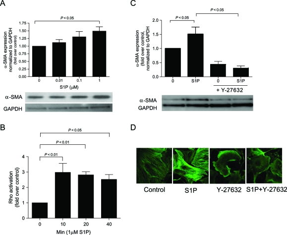
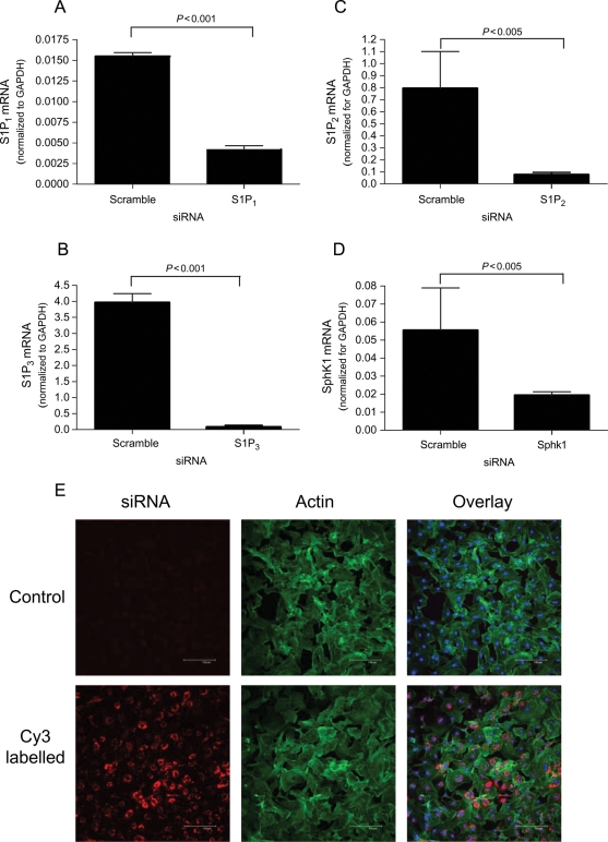
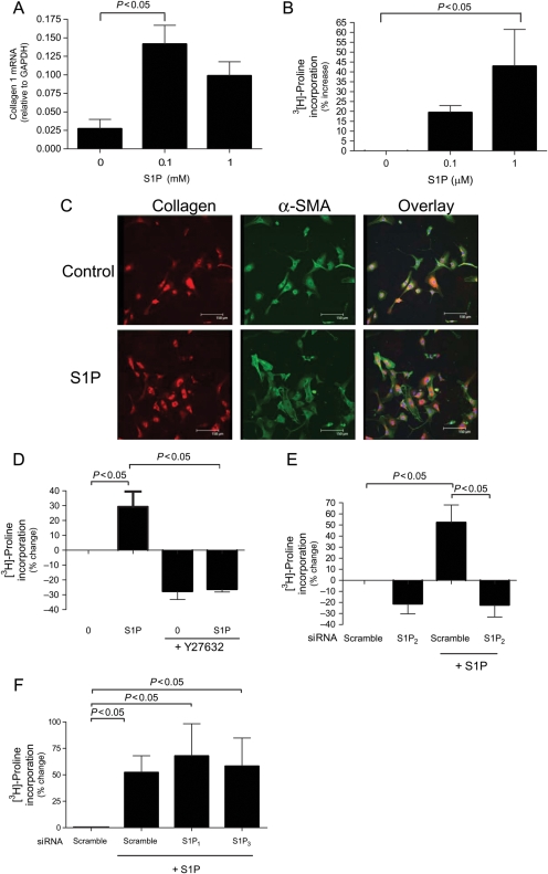
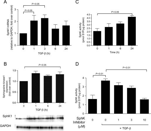
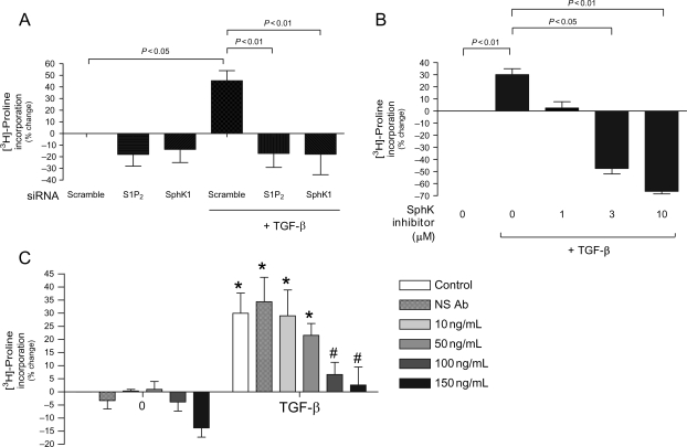
Methods and results: S1P-mediated effects on myofibroblast transformation, collagen production, and cross-talk with transforming growth factor-beta (TGF-beta) using mouse CF were examined. S1P increased alpha-smooth muscle actin (a myofibroblast marker) and collagen expression in a S1P2 receptor- and Rho kinase-dependent manner. TGF-beta increased sphingosine kinase 1 (SphK1; the enzyme responsible for S1P production) expression and activity. TGF-beta-stimulated collagen production was inhibited by SphK1 or S1P2 siRNA, a SphK inhibitor, and an anti-S1P monoclonal antibody.

Conclusion: These findings suggest that TGF-beta-stimulated collagen production in CF involves 'inside-out' S1P signalling whereby S1P produced intracellularly by SphK1 can be released and act in an autocrine/paracrine fashion to activate S1P2 and increase collagen production.


Figures

Figure 1
Figure 1
Sphingosine-1-phosphate (S1P)-stimulated myofibroblast transformation is Rho-dependent. α-SMA expression was measured using immunoblot or immunohistochemical analysis in cardiac fibroblasts that were (A) serum starved for 48 h and then stimulated for 48 h with serum-free media alone (control) or 0.01, 0.1, or 1 µM sphingosine-1-phosphate and (C and D) 1 µM sphingosine-1-phosphate with or without Y-27632 (Rho kinase inhibitor). (B) Rho activation was measured using Rho ELISA on cells that were serum starved for 48 h then stimulated with or without 1 µM sphingosine-1-phosphate for 10, 20 or 40 min. Data are represented as fold change over control. Values represent mean ± SEM of at least four independent experiments. Statistical analyses were performed using one-way analysis of variance with Bonferroni post hoc analysis.
Figure 2
Figure 2
siRNA transfection efficiency and mRNA knockdown. (AD) Quantitative polymerase chain reaction was used to evaluate mRNA of targeted genes after 48 h transfection with specific siRNAs or scramble siRNA. (E) Cardiac fibroblasts were transfected with Cy3 tagged scrambled siRNA or lipofectamine only (control) and imaged. Phalloidin (green) and Topro (blue) were used to label actin and nuclei, respectively. Data are represented as fold change over negative control siRNA only. Values represent mean ± SEM of at least three independent experiments. Statistical analyses were performed using Student’s t-test.
Figure 3
Figure 3
Sphingosine-1-phosphate (S1P)-stimulated myofibroblast transformation and Rho activation are S1P2-dependent. α-SMA expression was measured using immunoblot analysis in cardiac fibroblasts that were transfected with (A) S1P2 siRNA or (B) S1P1 or S1P3 siRNA for 48 h then treated for 48 h with serum-free media alone or 1 µM sphingosine-1-phosphate. Rho activation was measured using Rho ELISA on cells that were transfected with (C) S1P2 siRNA or (B) S1P1 or S1P3 siRNA for 48 h then stimulated with or without 1 µM sphingosine-1-phosphate. Data are represented as fold change over control. Values represent mean ± SEM of at least four independent experiments. Statistical analyses were performed using one-way analysis of variance with Bonferroni post hoc analysis.
Figure 4
Figure 4
Sphingosine-1-phosphate (S1P) increases collagen production in a Rho-dependent manner in cardiac fibroblast (CF). (A) Cardiac fibroblasts were serum starved for 48 h and then stimulated for 48 h with serum-free media alone (control) or 0.1, or 1 µM sphingosine-1-phosphate and collagen mRNA was measured by quantitative polymerase chain reaction using gene-specific primers. (C) Cardiac fibroblasts were serum starved for 48 h and then stimulated for 48 h with serum-free media alone (control) or 1 µM sphingosine-1-phosphate and collagen (red) and α-SMA (green) were measured using immunofluorescence and co-stained with Topro for nuclei (blue). Collagen expression was measured using collagenase sensitive [3H]-proline incorporation assay in cells that were (B) serum starved for 48 h and treated with serum-free media alone (control) or 0.1, or 1 µM sphingosine-1-phosphate (D) serum starved for 48 h followed by treatment with serum-free media alone or 1 µM sphingosine-1-phosphate with or without Y-27632 (Rho kinase inhibitor) and (E and F) transfected with S1P1, S1P2, or S1P3 siRNA for 48 h then treatment with or without 1 µM sphingosine-1-phosphate. Data are represented as percentage change from control. Values represent mean ± SEM of at least three independent experiments. Statistical analyses were performed using one-way analysis of variance with Bonferroni post hoc analysis.
Figure 5
Figure 5
Transforming growth factor-β (TGF-β) stimulates sphingosine kinase 1 (SphK1) mRNA, expression, and activity. Cardiac fibroblasts were serum starved for 48 h, treated with transforming growth factor-β (10 ng/mL) for the indicated times and (A) mRNA was harvested and evaluated for sphingosine kinase 1 expression, (B) protein was harvested and evaluated for sphingosine kinase 1 expression, (C) cell lysates were subjected to sphingosine kinase activity assay, and (D) cells were pre-treated for 30 min with various sphingosine kinase inhibitor concentrations followed by 24 h of transforming growth factor-β treatment. Data are represented as fold change over control or picomoles of sphingosine-1-phosphate/microgram protein. Values represent mean ± SEM of at least four independent experiments. Statistical analyses were performed using one-way analysis of variance with Bonferroni post hoc analysis.
Figure 6
Figure 6
Transforming growth factor-β (TGF-β-stimulated collagen expression is sphingosine kinase 1 (SphK1)- and S1P2-dependent. Collagen expression was measured using collagenase sensitive [3H]-proline incorporation assay in cells that were (A) transfected for 48 h with scramble control, S1P2, or sphingosine kinase 1 siRNA and then treated with serum-free media alone (control) or 10 ng/mL transforming growth factor-β (B) serum starved for 48 h followed by treatment with serum-free media alone or 10 ng/mL transforming growth factor-β with varying concentrations of sphingosine kinase inhibitor and (C) serum starved for 48 h followed by treatment with serum-free media alone or 10 ng/mL transforming growth factor-β with or without varying concentrations of anti-sphingosine-1-phosphate monoclonal antibody (S1P Ab) or non-specific (NS) Ab. Data are represented as percentage change from control. Values represent mean ± SEM of at least four independent experiments. Asterisk indicates significance from comparable treatment without transforming growth factor-β. Hash (#) indicates significant reduction from transforming growth factor-β without anti-sphingosine-1-phosphate monoclonal antibody. Statistical analyses were performed using one-way analysis of variance with Bonferroni post hoc analysis.

References

    1. Hinz B, Phan SH, Thannickal VJ, Galli A, Bochaton-Piallat ML, Gabbiani G. The myofibroblast: one function, multiple origins. Am J Pathol. 2007;170:1807–1816. - PMC - PubMed
    1. Banerjee I, Fuseler JW, Price RL, Borg TK, Baudino TA. Determination of cell types and numbers during cardiac development in the neonatal and adult rat and mouse. Am J Physiol Heart Circ Physiol. 2007;293:H1883–H1891. - PubMed
    1. Brown RD, Ambler SK, Mitchell MD, Long CS. The cardiac fibroblast: therapeutic target in myocardial remodeling and failure. Annu Rev Pharmacol Toxicol. 2005;45:657–687. - PubMed
    1. Sun Y, Kiani MF, Postlethwaite AE, Weber KT. Infarct scar as living tissue. Basic Res Cardiol. 2002;97:343–347. - PubMed
    1. Long CS, Brown RD. The cardiac fibroblast, another therapeutic target for mending the broken heart? J Mol Cell Cardiol. 2002;34:1273. - PubMed

Publication types

MeSH terms