

Curcumin inhibits prosurvival pathways in chronic lymphocytic leukemia B cells and may overcome their stromal protection in combination with EGCG
- PMID: 19228728
- PMCID: PMC3893060
- DOI: 10.1158/1078-0432.CCR-08-1511
Curcumin inhibits prosurvival pathways in chronic lymphocytic leukemia B cells and may overcome their stromal protection in combination with EGCG
Abstract
Purpose: Chronic lymphocytic leukemia (CLL) is incurable with current chemotherapy treatments. Curcumin (diferuloylmethane), an active ingredient in the spice turmeric, inhibits tumor metastasis, invasion, and angiogenesis in tumor cell lines. We evaluated the effects of curcumin on the viability of primary CLL B cells and its ability to overcome stromal mediated protection.
Experimental design: The in vitro effect of curcumin on primary CLL B cells was evaluated using fluorescence activated cell sorter analysis and Western blotting. For some experiments, CLL B cells were cocultured with human stromal cells to evaluate the effects of curcumin on leukemia cells cultured in their microenvironment. Finally, the effect of curcumin in combination with the green tea extract epigallocatechin-3 gallate (EGCG) was evaluated.
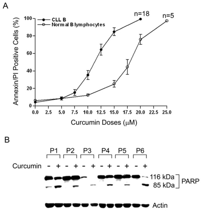
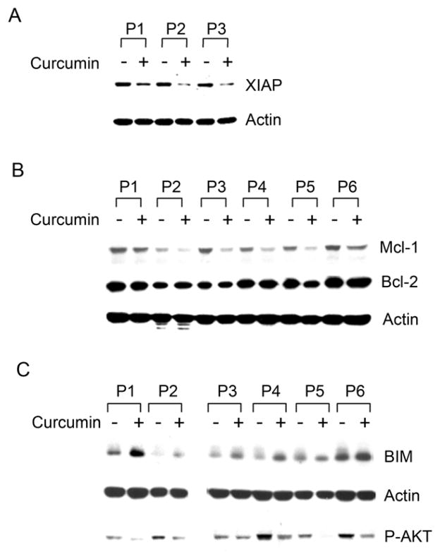
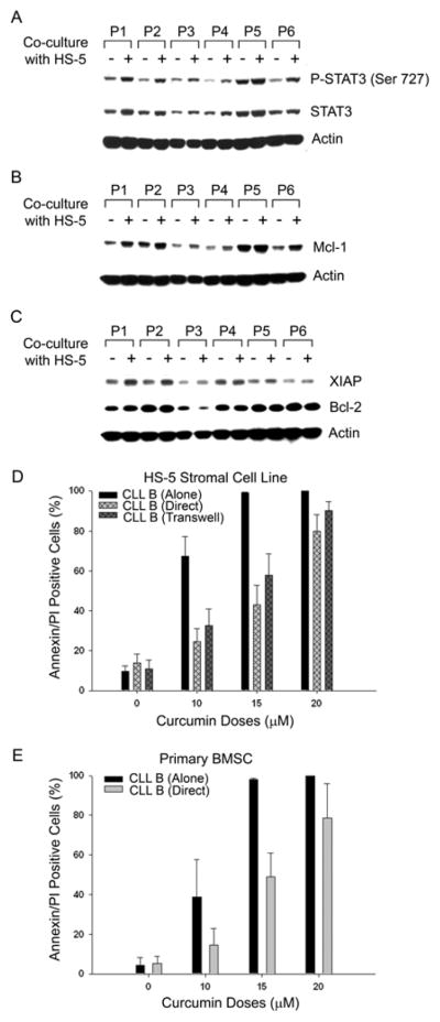
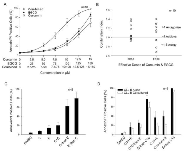
Results: Curcumin induced apoptosis in CLL B cells in a dose-dependent (5-20 micromol/L) manner and inhibited constitutively active prosurvival pathways, including signal transducers and activators of transcription 3 (STAT3), AKT, and nuclear factor kappaB. Moreover, curcumin suppressed expression of the anti-apoptotic proteins Mcl-1 and X-linked inhibitor of apoptosis protein (XIAP), and up-regulated the pro-apoptotic protein BIM. Coculture of CLL B cells with stromal cells resulted in elevated levels of STAT3, increased expression of Mcl-1 and XIAP, and decreased sensitivity to curcumin. When curcumin was administered simultaneously with EGCG, antagonism was observed for most patient samples. In contrast, sequential administration of these agents led to substantial increases in CLL B-cell death and could overcome stromal protection.
Conclusions: Curcumin treatment was able to overcome stromal protection of CLL B cells on in vitro testing and to synergize with EGCG when administered in a sequential fashion. Additional evaluation of curcumin as a potential therapeutic agent for treatment of CLL seems warranted.
Conflict of interest statement
Figures

Comment in
-
Turmeric and green tea: a recipe for the treatment of B-chronic lymphocytic leukemia.Clin Cancer Res. 2009 Feb 15;15(4):1123-5. doi: 10.1158/1078-0432.CCR-08-2791. Clin Cancer Res. 2009. PMID: 19228716 Free PMC article.
Similar articles
-
Bone marrow stroma-induced resistance of chronic lymphocytic leukemia cells to arsenic trioxide involves Mcl-1 upregulation and is overcome by inhibiting the PI3Kδ or PKCβ signaling pathways.Oncotarget. 2015 Dec 29;6(42):44832-48. doi: 10.18632/oncotarget.6265. Oncotarget. 2015. PMID: 26540567 Free PMC article.
-
Inhibition of NF-κB-mediated signaling by the cyclin-dependent kinase inhibitor CR8 overcomes prosurvival stimuli to induce apoptosis in chronic lymphocytic leukemia cells.Clin Cancer Res. 2013 May 1;19(9):2393-405. doi: 10.1158/1078-0432.CCR-12-2170. Epub 2013 Mar 26. Clin Cancer Res. 2013. PMID: 23532892
-
VEGF receptor phosphorylation status and apoptosis is modulated by a green tea component, epigallocatechin-3-gallate (EGCG), in B-cell chronic lymphocytic leukemia.Blood. 2004 Aug 1;104(3):788-94. doi: 10.1182/blood-2003-08-2763. Epub 2004 Mar 2. Blood. 2004. PMID: 14996703
-
Inflammation and survival pathways: chronic lymphocytic leukemia as a model system.Biochem Pharmacol. 2010 Dec 15;80(12):1936-45. doi: 10.1016/j.bcp.2010.07.039. Epub 2010 Aug 7. Biochem Pharmacol. 2010. PMID: 20696142 Free PMC article. Review.
-
NF-kB and the CLL microenvironment.Front Oncol. 2023 Mar 30;13:1169397. doi: 10.3389/fonc.2023.1169397. eCollection 2023. Front Oncol. 2023. PMID: 37064123 Free PMC article. Review.
Cited by
-
Polyphenols: Chemoprevention and therapeutic potentials in hematological malignancies.Front Nutr. 2022 Oct 26;9:1008893. doi: 10.3389/fnut.2022.1008893. eCollection 2022. Front Nutr. 2022. PMID: 36386899 Free PMC article. Review.
-
Molecular Action of Polyphenols in Leukaemia and Their Therapeutic Potential.Int J Mol Sci. 2021 Mar 17;22(6):3085. doi: 10.3390/ijms22063085. Int J Mol Sci. 2021. PMID: 33802972 Free PMC article. Review.
-
Evidence for and Against Green Tea and Turmeric in the Management of Chronic Lymphocytic Leukemia.Clin Lymphoma Myeloma Leuk. 2018 Oct;18(10):e421-e426. doi: 10.1016/j.clml.2018.06.021. Epub 2018 Jun 27. Clin Lymphoma Myeloma Leuk. 2018. PMID: 30007569 Free PMC article. Review.
-
Curcumin Combined with Thalidomide Reduces Expression of STAT3 and Bcl-xL, Leading to Apoptosis in Acute Myeloid Leukemia Cell Lines.Drug Des Devel Ther. 2020 Jan 15;14:185-194. doi: 10.2147/DDDT.S228610. eCollection 2020. Drug Des Devel Ther. 2020. PMID: 32021103 Free PMC article.
-
Role of the Dietary Phytochemical Curcumin in Targeting Cancer Cell Signalling Pathways.Plants (Basel). 2023 Apr 26;12(9):1782. doi: 10.3390/plants12091782. Plants (Basel). 2023. PMID: 37176840 Free PMC article. Review.
References
-
- Keating MJ. Chronic lymphocytic leukemia [Review] [65 refs] Seminars in Oncology. 1999;26:107–14. - PubMed
-
- Reed JC. Molecular biology of chronic lymphocytic leukemia. Semin Oncol. 1998;25:11–8. - PubMed
-
- Thomas A, El Rouby S, Reed JC, et al. Drug-induced apoptosis in B-cell chronic lymphocytic leukemia: relationship between p53 gene mutation and bcl-2/bax proteins in drug resistance. Oncogene. 1996;12:1055–62. - PubMed
-
- McConkey DJ, Chandra J, Wright S, et al. Apoptosis sensitivity in chronic lymphocytic leukemia is determined by endogenous endonuclease content and relative expression of BCL-2 and BAX. J Immunol. 1996;156:2624–30. - PubMed
Publication types
MeSH terms
Substances
Grants and funding
LinkOut - more resources
Full Text Sources
Miscellaneous
