. 2009 May 1;18(9):1600-11.
doi: 10.1093/hmg/ddp076. Epub 2009 Feb 19.

Delivery of bifunctional RNAs that target an intronic repressor and increase SMN levels in an animal model of spinal muscular atrophy

Affiliations

Delivery of bifunctional RNAs that target an intronic repressor and increase SMN levels in an animal model of spinal muscular atrophy

Travis D Baughan et al. Hum Mol Genet. .

Abstract

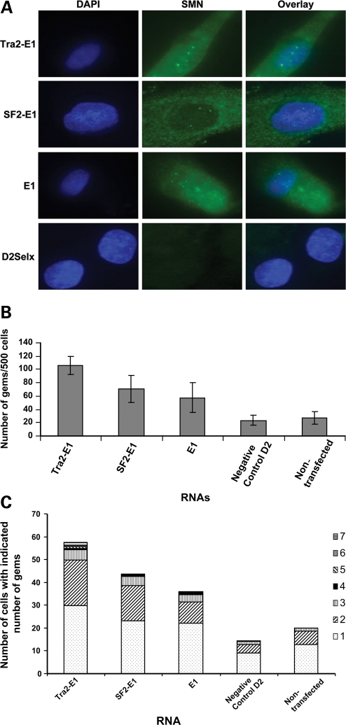
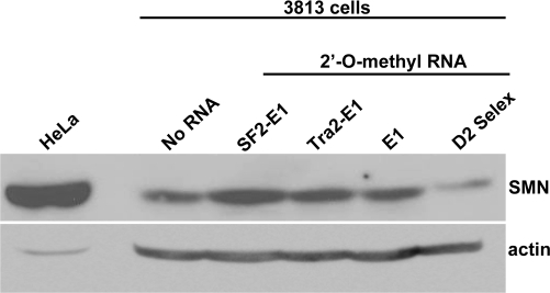
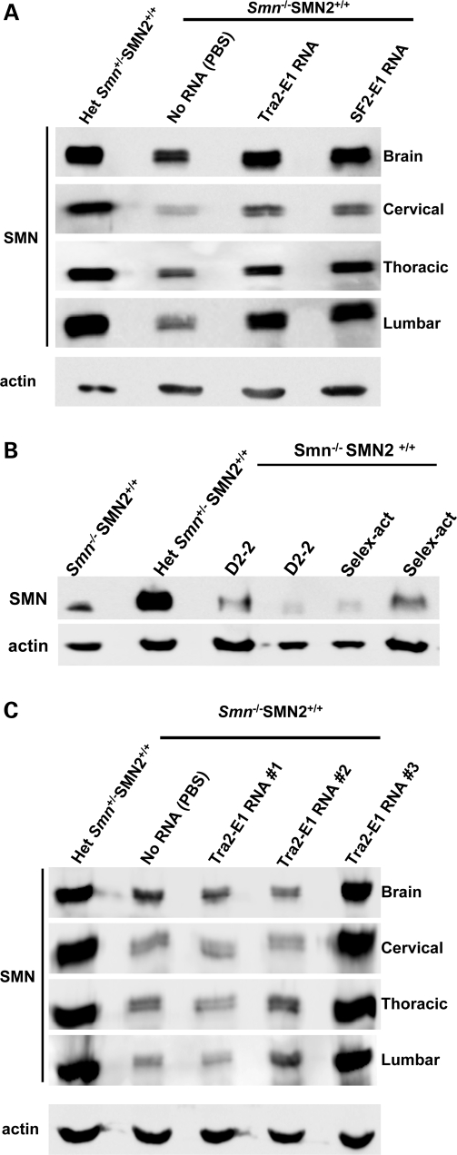
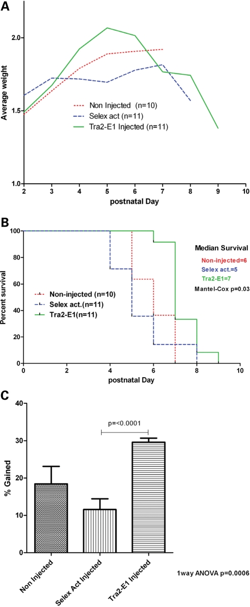
Spinal muscular atrophy (SMA) is a motor neuron disease caused by the loss of survival motor neuron-1 (SMN1). A nearly identical copy gene, SMN2, is present in all SMA patients, which produces low levels of functional protein. Although the SMN2 coding sequence has the potential to produce normal, full-length SMN, approximately 90% of SMN2-derived transcripts are alternatively spliced and encode a truncated protein lacking the final coding exon (exon 7). SMN2, however, is an excellent therapeutic target. Previously, we developed bifunctional RNAs that bound SMN exon 7 and modulated SMN2 splicing. To optimize the efficiency of the bifunctional RNAs, a different antisense target was required. To this end, we genetically verified the identity of a putative intronic repressor and developed bifunctional RNAs that target this sequence. Consequently, there is a 2-fold mechanism of SMN induction: inhibition of the intronic repressor and recruitment of SR proteins via the SR recruitment sequence of the bifunctional RNA. The bifunctional RNAs effectively increased SMN in human primary SMA fibroblasts. Lead candidates were synthesized as 2'-O-methyl RNAs and were directly injected in the central nervous system of SMA mice. Single-RNA injections were able to illicit a robust induction of SMN protein in the brain and throughout the spinal column of neonatal SMA mice. In a severe model of SMA, mean life span was extended following the delivery of bifunctional RNAs. This technology has direct implications for the development of an SMA therapy, but also lends itself to a multitude of diseases caused by aberrant pre-mRNA splicing.


Figures

Figure 1.
Figure 1.
E1 is a negative regulator of SMN 2 exon 7 inclusion. (A) RT–PCR of SMN minigenes (1 µg) transfected into HeLa cells, and total RNA was isolated after 24 h of transfection. Bands of exon 7 and Δ7 are shown by the use of previously published SMN minigene-specific primers. (B) Quantification of RT–PCR performed in triplicate on transfected cells utilizing a Cy3 fluorescent primer pair specific to SMN minigene; independent experiments repeated three times. This quantification shows an ∼2-fold increase of splicing to exon 7 when E1 is deleted. P-value ≤ 0.009.
Figure 2.
Figure 2.
Identification of PTB and FUSE-BP proteins bound to E1. (A) RNA affinity chromatography with subsequent Coommassie blue stain identified at least two specific proteins interacting with E1 but not with control RNA. The indicated bands were excised from the gel, and MALDI-TOF identified PTB and FUSE-BP as the two unique bands. (B) Confirmation of PTB interaction with E1. RNA-protein affinity chromatography was performed, and the western blot was developed using an anti-PTB-specific antibody. PTB specifically interacts with E1 RNA and not with the negative control RNA.
Figure 3.
Figure 3.
Schematic of E1-specific bifunctional RNAs. The organization of the bifunctional RNA is illustrated with the split antisense domain targeting the sequences flanking E1, with the non-specific linker sequence and three tandem repeats of high-affinity exonic splice enhancer sequences for either hTra2β1 or SF2/ASF. In addition, the schematic indicates the orientation of the U7-sm-opt sequence found on the plasmid-derived bifunctional RNAs.
Figure 4.
Figure 4.
Increase in SMN protein in the presence of plasmid expressed bifunctional RNAs. (A) SMA type 1 fibroblasts (3813 cells) were transiently transfected with 1 µg of plasmid DNA producing the indicated RNAs. Cells were incubated for 48 h and an immunoflourescence was performed. Unless indicated as the CMV promoter, the U6 promoter is driving RNA expression. Transfected cells are identified by GFP expression. Pictures are of representative cells found for each sample. (B) 3813 cells transfected with plasmids were randomly selected and gem numbers compiled. A total of 100 GFP-positive cells were observed and SMN-positive foci in the nucleus were counted (n = 3 and error bars are ±STD). (C) Gem data compiled and now expressed as the number of SMN-positive gems per nucleus (n = 3).
Figure 5.
Figure 5.
Increase in SMN expression after the transfection of 2′-O-methyl-modified bifunctional RNAs. (A) SMA type 1 fibroblasts (3813 cells) were transiently transfected with 100 ng of 2′-O-methyl bifunctional RNAs for 48 h. Immunoflourescence staining was performed and images are of representative cells. (B) Five hundred 3813 cells were randomly counted (n = 4) and the total gem number shown (error bars are ±STD). (C) Gem data compiled and expressed as the number of gems per nucleus (n = 4).
Figure 6.
Figure 6.
2′-O-methyl bifunctional RNAs increased total SMN protein levels in SMA fibroblasts. Subconfluent 3813 cells were transfected with 100 ng of the indicated RNAs and subsequently incubated for 48 h. Relative SMN protein levels were determined by western blot. HeLa cellular extracts were included as a size control.
Figure 7.
Figure 7.
ICV injection of 2′-O-methyl bifunctional RNAs increases SMN protein levels throughout the CNS. (A) Post-natal day (PND) 2 SMA mice were ICV-injected with 4 µg of the modified bifunctional RNAs, SF2-E1 and Tra2-E1 or PBS. Indicated tissues were isolated 24 h after injections. Western blots were done in quadruplicate, and a representative blot is shown. Multiple mice were injected and tested [Tra2-E1 (n = 13); SF2-E1 (n = 10), data not shown]. (B) Control RNAs do not elevate SMN protein levels. Four micrograms of modified control RNAs were delivered via ICV injection into PND 2 SMA mice and brain tissue was isolated 24 h post-injection. SMN protein levels were observed by western blot, which were done in triplicate, and a representative blot is shown. D2-2 and Selex-act are the two previously described RNAs, corresponding to an SMN intron 7 antisense (D2-2) or three tandem repeats of an in vitro affinity-determined binding motif for hnRNP-A1 (Selex-act). (C) ICV injection of Tra2-E1-modified bifunctional RNA increases SMN protein levels 5 days after a single injection. 2 PND SMA mice were ICV-injected with 4 µg of Tra2-E1 RNA. Five days following injection, the indicated tissues were harvested and SMN western blot performed. Western blots were done in quadruplicate and representative blot is shown.
Figure 8.
Figure 8.
ICV injection of 2′-O-methyl bifunctional RNA increases weight in a severe mouse model of SMA. (A) 2 PND severe mice were ICV-injected on PND 2, 4 with 6 µg of either Tra2-E1 or Selex activation RNA. Mice were weighed everyday post-first injection and graphed. (B) Kaplan–Meier survival curve depicts a trend towards increased life expectancy for Tra2-E1-injected mice (Mantel–Cox P = 0.03). (C) Percent weight gained post-first injection until maximal weight was graphed. Tra2-E1-injected mice showed a significant weight gain from birth to peak over non-injected and a negative control (Selex act) injected (one-way ANOVA P = 0.0006). There is a significant difference between Tra2-E1- and Selex act-injected mice (t-test P = < 0.0001).

References

    1. Crawford T.O., Pardo C.A. The neurobiology of childhood spinal muscular atrophy. Neurobiol. Dis. 1996;3:97–110. - PubMed
    1. Feldkotter M., Schwarzer V., Wirth R., Wienker T.F., Wirth B. Quantitative analyses of SMN1 and SMN2 based on real-time lightCycler PCR: fast and highly reliable carrier testing and prediction of severity of spinal muscular atrophy. Am. J. Hum. Genet. 2002;2:358–368. - PMC - PubMed
    1. Meister G., Fischer U. Assisted RNP assembly: SMN and PRMT5 complexes cooperate in the formation of spliceosomal UsnRNPs. EMBO J. 2002;21:5853–5863. - PMC - PubMed
    1. Lorson C.L., Hahnen E., Androphy E.J., Wirth B. A single nucleotide in the SMN gene regulates splicing and is responsible for spinal muscular atrophy. Proc. Natl Acad. Sci. USA. 1999;96:6307–6311. - PMC - PubMed
    1. Cartegni L., Krainer A.R. Disruption of an SF2/ASF-dependent exonic splicing enhancer in SMN2 causes spinal muscular atrophy in the absence of SMN1. Nat. Genet. 2002;30:377–384. - PubMed

Publication types

MeSH terms

Substances