. 2009 May;85(5):862-70.
doi: 10.1189/jlb.0408257. Epub 2009 Feb 19.

Murine dendritic cell antigen-presenting cell function is not altered by burn injury

Affiliations

Murine dendritic cell antigen-presenting cell function is not altered by burn injury

Satoshi Fujimi et al. J Leukoc Biol. 2009 May.

Abstract

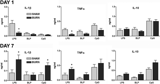
Severe injury disrupts normal immune regulation causing a transient hyperinflammatory reaction and suppressed adaptive immune function. This report addresses the potential contribution of dendritic cells (DC) to changes in adaptive immune function after injury by specifically measuring injury-induced changes in splenic DC numbers and subsets, cell-surface markers, TLR responses, and APC function. Using a mouse burn injury model, we found that injury did not markedly alter the relative percentage of lymphoid, myeloid, or plasmacytoid DC in the spleens of burn-injured mice. Moreover, we did not observe a significant reduction in cell-surface expression of several major costimulatory molecules, CD40, CD80, CD86, programmed death 1 ligand, ICOS ligand, and B7-H3, on DC. Instead, we observed increased cell-surface expression of CD86 at 1 day after injury with no significant changes in costimulatory molecule expression at 7 days after injury, suggesting that burn injury causes an early activation of DC. In addition, injury did not suppress DC reactivity to TLR2, TLR4, or TLR9 agonists. Most important, DC prepared from injured mice were able to present peptide antigen to naive OTII TCR transgenic CD4+ T cells as efficiently and effectively as DC from sham-injured mice. We also found that CD4 T cells stimulated with antigen presented by DC from sham or burn mice showed similar levels of IL-2, IFN-gamma, IL-10, and IL-13 production. Taken together, these findings support the conclusion that DC do not acquire a suppressive phenotype following severe injury in mice.


Figures

Fig. 1.
Fig. 1.
Injury-induced changes in DC numbers and subsets. At Days 1 and 7 after sham or burn injury, spleen cells were harvested from mice and then stained with CD11c antibody to identify DC and anti-CD8, anti-CD4, or anti-B220 antibody to identify lymphoid (CD8+), myeloid (CD4+), or plasmacytoid (B220+) DC subsets. The data shown represent the mean ± sem of n = 7–11 mice from two experiments. *, P < 0.05, sham versus burn by ANOVA.
Fig. 2.
Fig. 2.
Changes in costimulatory molecule expression on DC at 1 and 7 days after injury. Spleen cells were prepared from individual mice at Day 1 or 7 after burn injury. The cell suspensions were stained with FITC-labeled anti-CD11c antibody and counterstained with the indicated PE-labeled costimulatory molecule-specific antibodies. (A) Representative FACS plots showing costimulatory molecule-staining levels on gated CD11c+ cells; the numbers in the upper-right quadrants represent the percent-positive staining cells. (B) The plot shows the mean ± sem of n = 5–6 mice per group and is representative of three independent experiments. *, P < 0.05, sham versus burn by ANOVA.
Fig. 3.
Fig. 3.
TLR2-, TLR4-, and TLR9-induced effects on costimulatory molecule expression on DCs at 1 and 7 days after injury. Spleen cells were prepared from individual mice at Day 1 or 7 after burn injury. Purified CD11c+ cells were plated at 1 × 10⁵ per well of a 96-well plate and stimulated with 1 μg/ml or BLP, LPS, or CpG ODN 1826. After 18 h incubation, cell suspensions were stained with FITC-labeled anti-CD11c antibody and counterstained with the indicated PE-labeled, anticostimulatory molecule antibodies. The data represent the mean ± sem of n = 3 mice per group of three independent experiments. *, P < 0.05, sham versus burn by ANOVA.
Fig. 4.
Fig. 4.
The influence of injury on TLR2-, TLR4-, and TLR9-stimulated cytokine production by DC, which were purified from the spleens of sham and burn mice at Day 1 or 7 after injury and plated at a density of 1 × 10⁵ per well of a 96-well plate. After 48 h stimulation with 1 μg/ml or BLP, LPS, or CpG ODN 1826, supernatants were harvested and tested for cytokine production by ELISA. The data presented represent the mean ± sem of n = 3 mice per group of three independent experiments. *, P < 0.05, sham versus burn by ANOVA.
Fig. 5.
Fig. 5.
Injury-induced effect on DC APC activity. At 1 or 7 days after injury, DC were purified from the spleens of sham and burn mice and plated at a density of 5 × 10⁴ per well of a 96-well plate. CD4 T cells purified from OTII TCR transgenic mice (2×10⁵ cells) were cultured with DC and OVA323–339 peptide (1 μg/ml). After 48 h stimulation, supernatants were harvested and tested for cytokine production by ELISA. We did not detect any cytokine production when peptide was added to cultures of purified CD4 T cells from OTII mice in the absence of DC. The data represent the mean ± sem of four to six independent experiments. There was no significant difference between groups (P<0.05) as determined by ANOVA.
Fig. 6.
Fig. 6.
The influence of TLR stimulation on DC APC function. DC were purified from the spleens of sham and burn mice and plated at a density of 5 × 10⁴ per well of a 96-well plate with CD4 T cells purified from OTII TCR transgenic mice (2×10⁵ cells) and OVA323–339 peptide (1 μg/ml). LPS, BLP, or CpG ODN (1 μg/ml) were added to the cultures, and after 48 h incubation, supernatants were harvested to test for cytokine production by ELISA. The data represent the mean ± sem of five independent experiments. *, P < 0.05, for sham versus sham; **, P < 0.05, for burn versus burn between TLR agonists as determined by ANOVA. There was no significant difference between sham and burn groups (P<0.05) as determined by ANOVA.

References

    1. Smith J W, Gamelli R L, Jones S B, Shankar R. Immunologic responses to critical injury and sepsis. J Intensive Care Med. 2006;21:160–172. - PubMed
    1. Wilkinson R A, Fishman J A. Effect of thermal injury with Pseudomonas aeruginosa infection on pulmonary and systemic bacterial clearance. J Trauma. 1999;47:912–917. - PubMed
    1. Kobayashi M, Kobayashi H, Herndon D N, Pollard R B, Suzuki F. Burn-associated Candida albicans infection caused by CD30+ type 2 T cells. J Leukoc Biol. 1998;63:723–731. - PubMed
    1. Kobayashi H, Kobayashi M, Utsunomiya T, Herndon D N, Pollard R B, Suzuki F. Therapeutic protective effects of IL-12 combined with soluble IL-4 receptor against established infections of herpes simplex virus type 1 in thermally injured mice. J Immunol. 1999;162:7148–7154. - PubMed
    1. Zedler S, Bone R C, Baue A E, von Donnersmarck G H, Faist E. T-cell reactivity and its predictive role in immunosuppression after burns. Crit Care Med. 1999;27:66–72. - PubMed

Publication types