. 2009 Mar 10;106(10):3883-8.
doi: 10.1073/pnas.0813334106. Epub 2009 Feb 19.

IL-32-dependent effects of IL-1beta on endothelial cell functions

Affiliations

IL-32-dependent effects of IL-1beta on endothelial cell functions

Claudia A Nold-Petry et al. Proc Natl Acad Sci U S A. .

Abstract

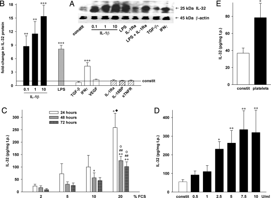
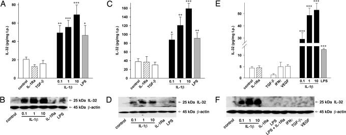
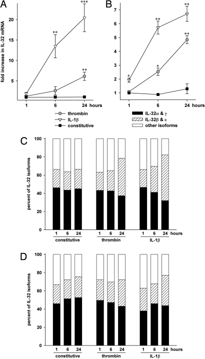
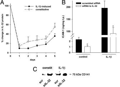
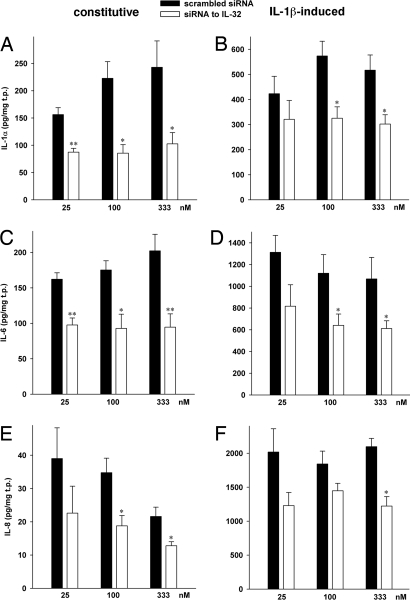
Increasing evidence demonstrates that interleukin (IL)-32 is a pro-inflammatory cytokine, inducing IL-1alpha, IL-1beta, IL-6, tumor necrosis factor (TNF)-alpha, and chemokines via nuclear factor (NF)-kappaB, p38 mitogen-activated protein kinase (MAPK), and activating protein (AP)-1 activation. Here we report that IL-32 is expressed and is also functional in human vascular endothelial cells (EC) of various origins. Compared with primary blood monocytes, high levels of IL-32 are constitutively produced in human umbilical vein EC (HUVEC), aortic macrovascular EC, and cardiac as well as pulmonary microvascular EC. At concentrations as low as 0.1 ng/ml, IL-1beta stimulated IL-32 up to 15-fold over constitutive levels, whereas 10 ng/ml of TNFalpha or 100 ng/ml of lipopolysaccharide (LPS) were required to induce similar quantities of IL-32. IL-1beta-induced IL-32 was reduced by inhibition of the IkappaB kinase-beta/NF-kappaB and ERK pathways. In addition to IL-1beta, pro-coagulant concentrations of thrombin or fresh platelets increased IL-32 protein up to 6-fold. IL-1beta and thrombin induced an isoform-switch in steady-state mRNA levels from IL-32alpha/gamma to beta/epsilon. Adult EC responded in a similar fashion. To prove functionality, we silenced endogenous IL-32 with siRNA, decreasing intracellular IL-32 protein levels by 86%. The knockdown of IL-32 resulted in reduction of constitutive as well as IL-1beta-induced intercellular adhesion molecule-1 (ICAM-1) (of 55% and 54%, respectively), IL-1alpha (of 62% and 43%), IL-6 (of 53% and 43%), and IL-8 (of 46% and 42%). In contrast, the anti-inflammatory/anti-coagulant CD141/thrombomodulin increased markedly when IL-32 was silenced. This study introduces IL-32 as a critical regulator of endothelial function, expanding the properties of this cytokine relevant to coagulation, endothelial inflammation, and atherosclerosis.


Conflict of interest statement

The authors declare no conflict of interest.

Figures

Fig. 1.
Fig. 1.
Expression and regulation of IL-32 in HUVEC. HUVEC were incubated with the indicated stimuli and/or inhibitors for 20 hours (A, B, D, E) or for the periods indicated (C). Concentrations (in ng/ml) were as follows: LPS, 100; TGF-β1, 20; IFNγ, 25; VEGF, 25; IL-1Ra, 10000; IL-18BP, 1000; sTNFR, 10000. (B) Mean fold-changes in IL-32 protein levels normalized to total protein (in mg) ± SEM; n = 9; *P < 0.05; **P < 0.01; and ***P < 0.001 for stimulated vs. untreated cultures. Normalized absolute IL-32 concentrations ranged from 28 to 269 pg/mg in controls and from 581 to 4025 pg/mg in IL-1β– (10 ng/ml) stimulated cells. (A) One representative of five independently performed Western blots of HUVEC cell lysates is shown. (C) HUVEC were cultured with different concentrations of FCS. Normalized absolute IL-32 levels ± SEM are shown; n = 5; *P < 0.05; and **P < 0.01 for 2% vs. 10 or 20%; ##P < 0.01 for 5% vs. 20%; oP < 0.05 for 10% vs. 20%; ♦, 20% at 24 hours vs. 20% at 72 hours. (D) IL-32 protein after incubation with the indicated concentrations of thrombin, mean ± SEM; n = 5; *P < 0.05; and **P < 0.01 for constitutive vs. thrombin. (E) Effect of incubation of HUVEC with 50 × 106 platelets on IL-32 is depicted. n = 3; *P < 0.05 for constitutive vs. platelets.
Fig. 2.
Fig. 2.
IL-32 production in EC of different origins. AEC (A, B), CMEC (C, D), and PMEC (E, F) were treated with the indicated stimuli (concentrations as in Fig. 1) or IL-1Ra (10 μg/ml) for 20 hours in the presence of 2% FCS. “Control” indicates constitutive production of IL-32. IL-1β concentrations are displayed in ng/ml. Cell lysates were harvested and IL-32 protein levels were determined. (A, C, E) The absolute IL-32 concentrations normalized to total protein are shown. Mean ± SEM; n = 4 for AEC and n = 8 for CMEC and PMEC; *P < 0.05; **P < 0.01; and ***P < 0.001 for treated cells vs. controls. (B, D, F) One of four blots resulting from independent experiments from each cell type is shown.
Fig. 3.
Fig. 3.
Expression and regulation of IL-32 mRNA isoforms in EC. Two hours after changing from growth- to stimulation medium, HUVEC (A, C) or CMEC (B, D) were stimulated with 10 ng/ml IL-1β or 7.5 U/ml thrombin for the indicated periods of time or were left untreated. Relative mRNA quantities were calculated using the ΔΔCT method. (A, B) Constitutive mRNA levels at 1 hour are defined as background. Mean fold increases ± SEM in all isoforms of IL-32 mRNA over background are shown; n = 4; *P < 0.05; **P < 0.01; and ***P < 0.001 for treated cells vs. controls. (C, D) The fractions of IL-32α/γ, β/ε, and other isoforms are depicted for each stimulation condition. The levels of all isoforms for each sample were set at 100%; n = 4.
Fig. 4.
Fig. 4.
Knockdown of IL-32 and its effect on ICAM-1 and thrombomodulin/CD141. (A) After transfection and stimulation with 10 ng/ml IL-1β, EC were cultured for the indicated time periods and IL-32 was measured in cell lysates. The graph depicts mean percent changes in IL-32 protein levels comparing siIL-32-transfected to scrambled-transfected cells ± SEM; n = 3. (B, C) HUVEC transfected with 100 nmol/l of either siIL-32 or scrambled were stimulated with 10 ng/ml IL-1β for 20 hours (B) or 30 minutes (C) or left untreated. (B) ICAM-1 was assayed by ELISA in cell lysates. Means of normalized protein concentration ± SEM in ng/mg is shown; n = 6; *P < 0.05 for siIL-32 compared with scrambled. (C) One representative of four Western blots of HUVEC lysates is depicted. The degree of silencing in these lysates is shown in Fig. S1 A and B.
Fig. 5.
Fig. 5.
The effects of siIL-32 on cytokine levels in HUVEC. Cell lysates (A, B) or supernatants (C–F) from unstimulated (Left column) or IL-1β-treated (10 ng/ml, Right column) HUVEC transfected with concentration-matched pairs of either siIL-32 or scrambled siRNA were analyzed for protein levels of IL-1α (A, B), IL-6 (C, D), and IL-8 (E, F) after a 20-hour incubation period. IL-32 levels in these cultures are shown in Fig. S1 A and B. Means of normalized absolute cytokine concentrations ± SEM are shown; n = 10; *P < 0.05; and **P < 0.01 for siIL-32 compared with scrambled.

Similar articles

Cited by

References

    1. Kim SH, et al. Interleukin-32: A cytokine and inducer of TNFalpha. Immunity. 2005;22:131–142. - PubMed
    1. Nold MF, et al. Endogenous IL-32 controls cytokine and HIV-1 production. J Immunol. 2008;181:557–565. - PubMed
    1. Goda C, et al. Involvement of IL-32 in activation-induced cell death in T cells. Int Immunol. 2006;18:233–240. - PubMed
    1. Netea MG, et al. Mycobacterium tuberculosis induces interleukin-32 production through a caspase-1/IL-18/interferon-gamma-dependent mechanism. PLoS Med. 2006;3:e277. - PMC - PubMed
    1. Shoda H, et al. Interactions between IL-32 and tumor necrosis factor alpha contribute to the exacerbation of immune-inflammatory diseases. Arthritis Res Ther. 2006;8:R166. - PMC - PubMed

Publication types

MeSH terms

LinkOut - more resources