. 2009 Mar 10;106(10):4012-7.
doi: 10.1073/pnas.0811698106. Epub 2009 Feb 19.

Oligomeric amyloid beta associates with postsynaptic densities and correlates with excitatory synapse loss near senile plaques

Affiliations

Oligomeric amyloid beta associates with postsynaptic densities and correlates with excitatory synapse loss near senile plaques

Robert M Koffie et al. Proc Natl Acad Sci U S A. .

Abstract

Synapse loss correlates with a cognitive decline in Alzheimer's disease (AD), but whether this is caused by fibrillar deposits known as senile plaques or soluble oligomeric forms of amyloid beta (Abeta) is controversial. By using array tomography, a technique that combines ultrathin sectioning of tissue with immunofluorescence, allowing precise quantification of small structures, such as synapses, we have tested the hypothesis that oligomeric Abeta surrounding plaques contributes to synapse loss in a mouse model of AD. We find that senile plaques are surrounded by a halo of oligomeric Abeta. Analysis of >14,000 synapses (represented by PSD95-stained excitatory synapses) shows that there is a 60% loss of excitatory synapses in the halo of oligomeric Abeta surrounding plaques and that the density increases to reach almost control levels in volumes further than 50 microm from a plaque in an approximately linear fashion (linear regression, r(2) = 0.9; P < 0.0001). Further, in transgenic cortex, microdeposits of oligomeric Abeta associate with a subset of excitatory synapses, which are significantly smaller than those not in contact with oligomeric Abeta. The proportion of excitatory synapses associated with Abeta correlates with decreasing density (correlation, -0.588; P < 0.0001). These data show that senile plaques are a potential reservoir of oligomeric Abeta, which colocalizes with the postsynaptic density and is associated with spine collapse, reconciling the apparently competing schools of thought of "plaque" vs. "oligomeric Abeta" as the synaptotoxic species in the brain of AD patients.


Conflict of interest statement

The authors declare no conflict of interest.

Figures

Fig. 1.
Fig. 1.
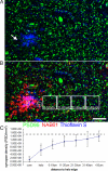
Oligomeric Aβ is present in the dense core of amyloid plaques and in a halo surrounding the core. (A) NAB61 antibody conjugated to Alexa Fluor 594 (red) applied topically in vivo shows oligomeric Aβ surrounding dense plaques (labeled with methoxy XO4; blue). (B) Postmortem staining of dense plaques by using methoxy XO4 (blue) and oligomeric Aβ by using NAB61 antibody (red) confirms that oligomeric Aβ is found in the core of plaques and in an area surrounding the core. (Scale bar: 10 μm.)
Fig. 2.
Fig. 2.
A 3D reconstruction from array tomography images of a 60 μm × 60 μm × 20 μm volume of APP/PS1 cortex shows that a large volume of cortex around the dense core of the plaque (ThioS; blue) is occupied by a halo of oligomeric Aβ (NAB61 staining; red). Further, the 3D reconstruction illustrates that postsynaptic densities (PSD95 stain; green) are lost both within the plaque core (arrow; A), and in the area surrounding the core (arrowhead; C), which is within the halo of oligomeric Aβ (B and D).
Fig. 3.
Fig. 3.
Precise quantification of PSD loss using array tomography. Postsynaptic densities (PSD95; green), oligomeric Aβ (NAB61; red), plaque cores (ThioS; blue), and nuclei (DAPI; blue) were stained on ribbons of 70-nm sections of APP/PS1 and nontransgenic cortex (part of a single section from the APP/PS1 cortex shown in A and B). To quantify the relationship between synapse loss and proximity to a plaque, images were taken of the same area on serial sections, the volumes aligned, and PSD95 puncta density was analyzed. Volumes were chosen within the plaque core (box C in B), the halo of oligomeric Aβ surrounding the core (arrow in A, box H in B), and in volumes extending out from the edge of the halo in 10-μm increments. Quantification (C) reveals significant PSD loss compared with control cortex (dotted line shows control average, ANOVA PSD density-dependent variable, genotype-independent F1,195 = 71.149, P < 0.0001). Density of PSDs also decreases in APP/PS1 cortex in areas near plaques (ANOVA synapse density-dependent variable, distance category independent split by genotype F6,123 = 36.323, P < 0.0001; synapse density correlates with average distance from halo edge correlation coefficient 0.706, P < 0.0001). *, P < 0.05 ANOVA split by distance category comparison to control average. (Scale bar: 10 μm.)
Fig. 4.
Fig. 4.
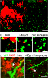
Oligomeric Aβ associates with PSDs. (A) Projection of images of array sections (500-nm total thickness) that have been processed by using the watershed analysis program (thresholded and puncta present in only one section excluded) show postsynaptic densities (green) and oligomeric Aβ (NAB61; red). (B) Higher-resolution projections from within the halo of oligomeric Aβ surrounding a plaque further than 50 μm from a plaque in APP/PS1 transgenic mouse cortex and a volume from nontransgenic cortex show that oligomeric Aβ staining is present at some postsynaptic densities (yellow). (C) Z-projections of raw images from an array of a mouse transgenic for YFP (stained green in C) and APP/PS1 demonstrate that NAB61 puncta (red) contact dendritic spines (arrows point to spine heads, yellow shows colocalization) both in the halo of oligomeric Aβ surrounding a plaque and distant from plaques. (Scale bars: A and C, 5 μm; B, 1 μm.
Fig. 5.
Fig. 5.
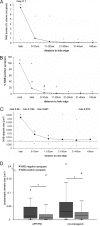
Quantification of oligomeric Aβ deposits and their association with PSDs. (A) The burden of oligomeric Aβ (percentage neuropil volume occupied by NAB61 staining) is increased in the APP/PS1 cortex compared with wild type (dotted line) (Mann–Whitney test P < 0.0001). The oligomeric Aβ burden also increases in volumes closer to plaques in APP/PS1 mice (Kruskal–Wallis test, distance-independent variable, split by genotype APP/PS1, P < 0.0001). (B) The percentage of excitatory synapses positive for oligomeric Aβ staining increases near plaques in the APP/PS1 trasngenic cortex (Kruskal–Wallis test, P < 0.0001). Post hoc tests show that the percentage of PSDs interacting with oligomeric Aβ is significantly higher than nontransgenic levels in the plaque core and halo, and there is a trend toward an increase between 0 and 10 μm away from the edge of the halo. (C) NAB61 deposits are larger in APP/PS1 mice than in wild-type cortex (Mann–Whitney test P < 0.0001), with very large deposits occurring, particularly in the halo (maximum size, 0.36 μm3). (D) A box plot of postsynaptic density size shows that PSDs are smaller in the APP/PS1 cortex than in nontransgenic cortex (Mann–Whitney test P < 0.0001), with a more substantial reduction in size in puncta associated with NAB61 deposits (Mann–Whitney test P = 0.0026). Even in nontransgenic brain, PSDs associated with oligomeric Aβ deposits are smaller than those not in contact with oligomeric Aβ (Mann–Whitney test P = 0.0243). Together, these data indicate that NAB61 interactions with the PSD may contribute to synapse loss. Data are not normally distributed and thus are presented as median with minima and maxima shown; control medians are dotted lines in A, B, and C. *, Post hoc Mann–Whitney tests P < 0.05; #, P = 0.06.

Similar articles

Cited by

References

    1. Terry RD, et al. Physical basis of cognitive alterations in Alzheimer's disease: Synapse loss is the major correlate of cognitive impairment. Ann Neurol. 1991;30:572–580. - PubMed
    1. DeKosky ST, Scheff SW, Styren SD. Structural correlates of cognition in dementia: Quantification and assessment of synapse change. Neurodegeneration. 1996;5:417–421. - PubMed
    1. Coleman PD, Yao PJ. Synaptic slaughter in Alzheimer's disease. Neurobiol Aging. 2003;24:1023–1027. - PubMed
    1. Shankar GM, et al. Amyloid-β protein dimers isolated directly from Alzheimer's brains impair synaptic plasticity and memory. Nat Med. 2008;14:837–842. - PMC - PubMed
    1. Snyder EM, et al. Regulation of NMDA receptor trafficking by amyloid-β. Nat Neurosci. 2005;8:1051–1058. - PubMed

Publication types

Substances

LinkOut - more resources