. 2009 Feb 18;29(7):2009-21.
doi: 10.1523/JNEUROSCI.5025-08.2009.

Involvement of the cytoplasmic C-terminal domain of connexin43 in neuronal migration

Affiliations

Involvement of the cytoplasmic C-terminal domain of connexin43 in neuronal migration

Cima Cina et al. J Neurosci. .

Abstract

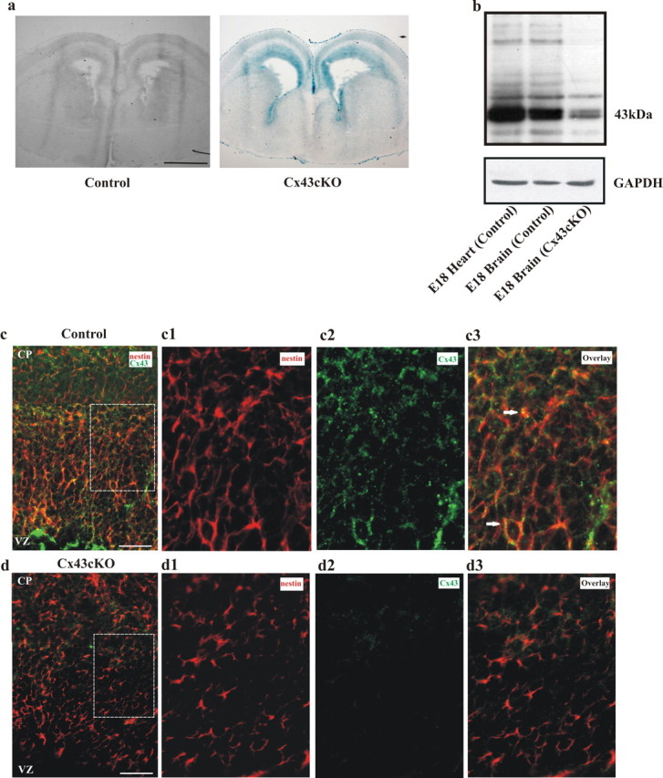
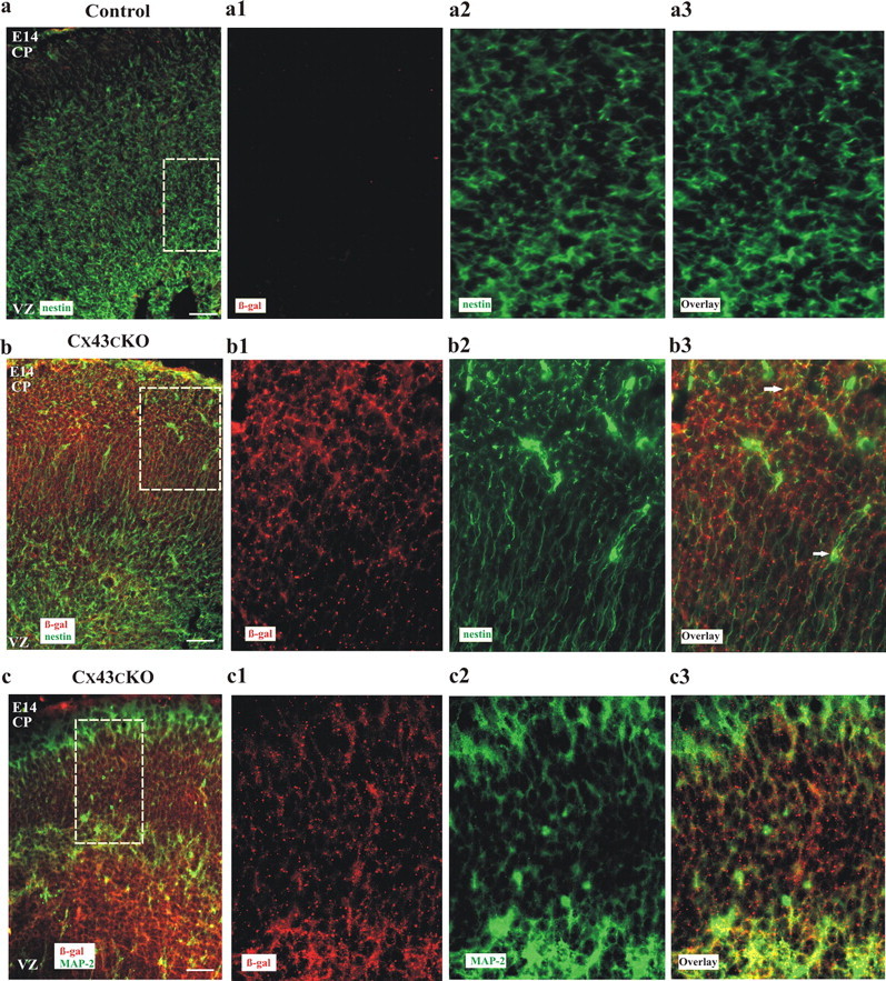
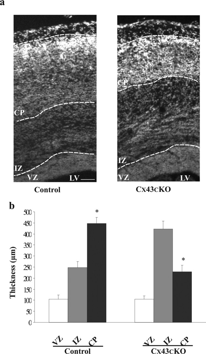
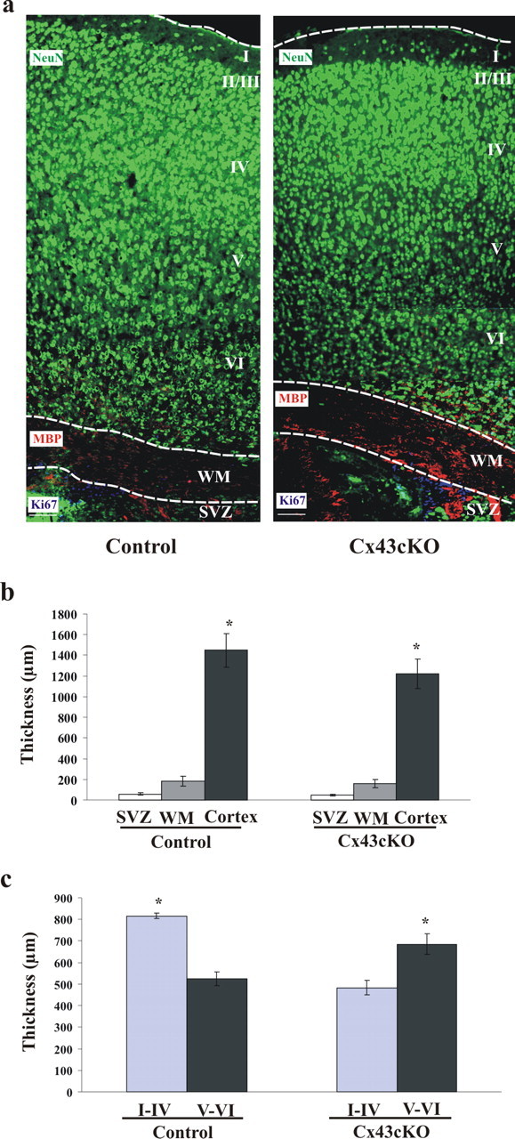
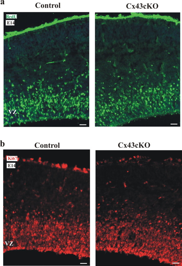
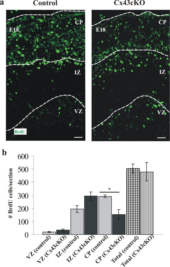
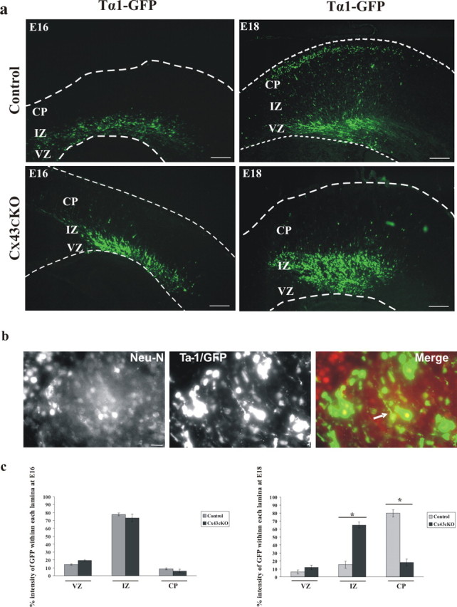
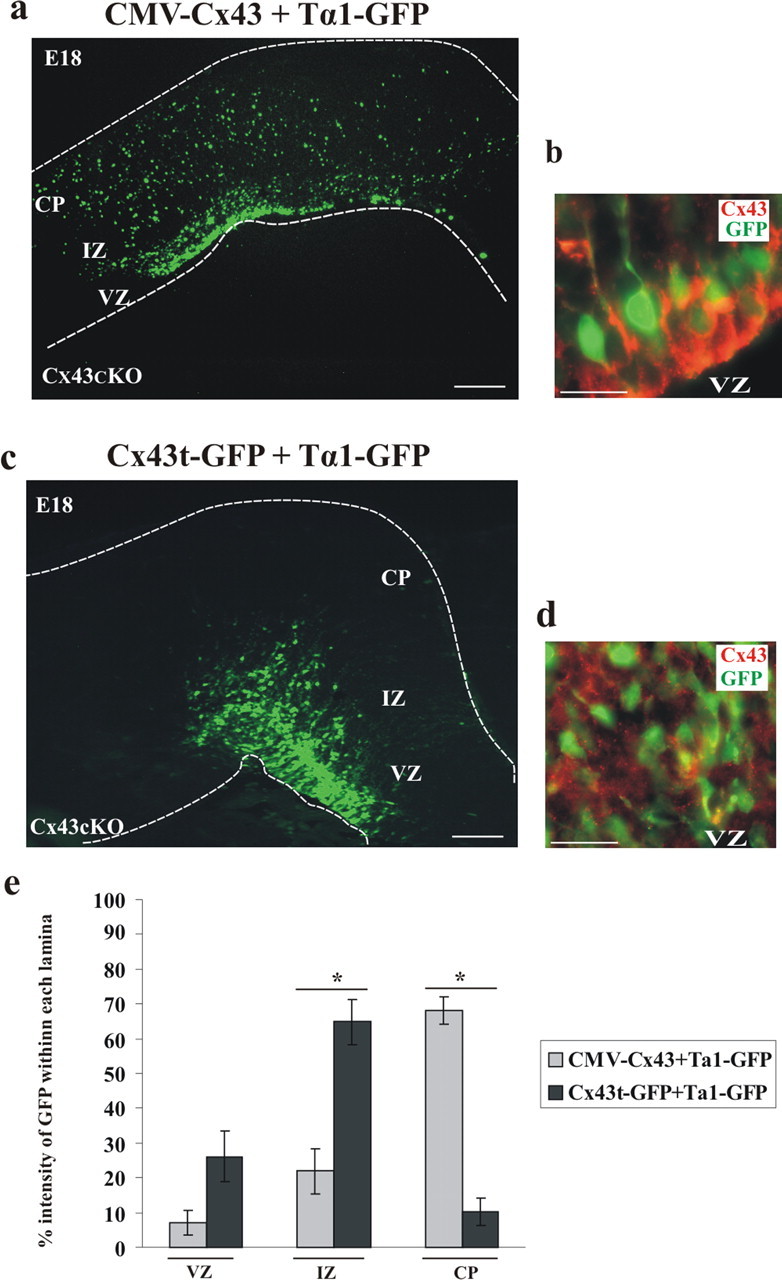
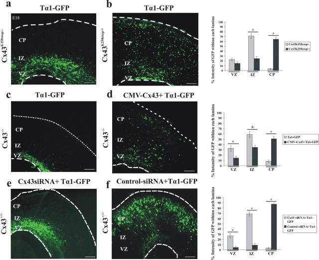
During brain development, young neurons closely associate with radial glial while migrating from the ventricular zone (VZ) to the cortical plate (CP) of the neocortex. It has been shown previously that gap junctions are needed for this migration to occur properly, but the precise mechanism responsible is still in question. Here, we used Cre recombinase, driven by the nestin promoter, to conditionally knock-out a floxed coding DNA of the connexin43 (Cx43) gene in mice. Radial glia in the VZ normally express connexin43. They undergo divisions that produce neurons and astrocytes and serve as migratory guides for the daughter cells that they produce. Based on histological analysis, we suggest that removing Cx43 from radial glia alters the normal lamination of the mouse neocortex. To monitor newborn neurons during development, we introduced a plasmid containing green fluorescent protein driven by a neuronal (Talpha1 tubulin) promoter into the embryonic neocortex using in utero electroporation. The transfected migrating neurons remain in the VZ/intermediate zone (IZ) of the Cx43 conditional knock-out (Cx43cKO) animals, whereas in Cx43(fl/fl) mice, neurons migrate through the IZ into the CP, indicating that deletion of Cx43 from nestin-positive cells disrupts neuronal migration. We were able to rescue migration of Cx43cKO neurons by electroporating a cytomegalovirus-Cx43 expression plasmid into the embryonic cortex. In contrast, a C-terminal truncated form of Cx43 failed to rescue neuronal migration. In addition, Cx43(K258stop) mice, in which Cx43 lacks the last 125 amino acid residues of the cytoplasmic C-terminal domain, gave results similar to those seen with the Cx43cKO mice. This study illustrates that deletion of the C-terminal domain of Cx43 alters neuronal migration in the neocortex.


Figures

Figure 1.
Figure 1.
Cre activity in Cx43fl/fl; nestin–Cre (Cx43cKO) mice. a, Coronal sections of E14 embryonic forebrain from control Cx43fl/fl (left) and Cx43cKO (right) mice were stained for lacZ, indicating Cre-mediated loss of Cx43 expression. Cre activity was observed in Cx43cKO mice but not in control mice. n = 4. b, Representative immunoblot showing the reduction of Cx43 protein in the neocortex of Cx43cKO mice. Equal amounts of embryonic heart and cortical tissue protein were immunoblotted and probed with antibodies recognizing Cx43 (43 kDa). GAPDH was used as a loading control. n = 3. c, d, Cx43cKO mice display a strong decrease in the level of Cx43 expression throughout the neocortex compared with control mice. Coronal brain sections of E18 littermates were stained for nestin and Cx43. Cx43 showed association (arrows) with nestin-expressing cells in control animals (c) and was absent in Cx43cKO sections (d). c1–d3 are higher-magnification micrographs of the areas outlined in c and d. n = 4. Scale bars: a, 1 mm; c, d, 100 μm; c1–d3, 40 μm.
Figure 2.
Figure 2.
Cre activity is observed in radial glia and MAP-2-positive neurons in Cx43cKO brains. Coronal sections of control (a) and Cx43cKO (b, c) neocortices were stained for β-gal (red), MAP-2, and/or nestin (green). Control brains were devoid of β-gal labeling compared with Cx43cKO littermates (a–c). a1–c3 represent higher-magnification micrographs of the section in a–c. Note that β-gal staining is associated with both nestin and MAP-2 expression in Cx43cKO mice. Arrows represent association of nestin- or MAP-2-expressing cells with β-gal. n = 3. Scale bars: a–c, 100 μm; a1–c3, 40 μm.
Figure 3.
Figure 3.
Deletion of Cx43 in the developing neocortex perturbs normal cortical lamination. a, Thickness of the VZ, IZ, and CP was measured in the neocortices of control and Cx43cKO mice. The cortical sections were stained with DAPI, and the cortical layers were identified based on their anatomical cell distribution. Using AxioVision software, the identified border lines between layers were labeled. In the cerebral cortices of E18 Cx43cKO mice, the thickness of the IZ was increased and the CP decreased compared with the IZ of control mice. b, The representative graph shows that the IZ in the Cx43cKO mice is increased in size compared with the control mice, whereas in control mice the CP is larger. *p < 0.05, Student's t test. n = 4. LV, Lateral ventricle. Scale bar, 100 μm.
Figure 4.
Figure 4.
Loss of Cx43 in the developing neocortex leads to a reduction in the thickness of the postnatal neocortex. a, Thickness of the SVZ, WM, and the cortex was measured in the P16 cortices of control and Cx43cKO mice. From each animal, 10 cortical sections spanning rostral end to the caudal end were stained for NeuN (green) to label mature neurons in the cortex (lamina I–VI), MBP (red) to label myelin fibers in the WM, and Ki67 (blue) to label proliferating cells in the SVZ. Using AxioVision software, the outline of each layer was drawn and the thickness was measured. b, The representative graph shows that, compared with the control littermates at P16, the cortex of the Cx43cKO mice was thinner. c, The representative graph shows significantly reduced thickness of upper cortical layers and increased thickness of lower cortical layers in Cx43cKO, but the normal layering is maintained. *p < 0.05, Mann–Whitney test. n = 5. Scale bars, 100 μm.
Figure 5.
Figure 5.
Deletion of Cx43 in radial glia of Cx43cKO mice does not produce a change in the rate of proliferation. Representative sections of BrdU labeling at E14 are shown in a. BrdU was administered at E14, and brains were fixed 2 h after labeling. Immunohistochemistry for BrdU was performed. From at least four brains per group, 10 coronal sections were assessed spanning the rostral to the caudal end of the neocortex. Based on DAPI staining, the sections of the neocortex were divided into three zones: the VZ, the IZ, and the CP. To define each zone, the outline of the VZ, the IZ, and the CP of each section was traced using the AxioVsion 4.2 software. Selected samples were also double labeled with polyclonal antibodies for Ki67 and showed a normal distribution of proliferative cells in the Cx43cKO mice (b). n = 4. Scale bars, 100 μm.
Figure 6.
Figure 6.
Loss of Cx43 in radial glia causes accumulation of BrdU+ cells in the VZ/IZ of Cx43cKO forebrains. a, Representative sections of BrdU labeling in E18 cortex of mice exposed to BrdU at E14. In Cx43cKO mice, there were still BrdU-labeled cells positioned in the VZ compared with the control mice. b, Distribution of BrdU-labeled cells in the IZ and the CP of the neocortex at E18. In the Cx43cKO mice, the number of BrdU-labeled cells in the CP was decreased and in the IZ increased compared with the control mice. Total number of cells in all zones was counted, and no significant difference was found among all the animals studied. *p < 0.05, Student's t test. n = 6. Scale bars, 100 μm.
Figure 7.
Figure 7.
Deletion of Cx43 in radial glial cells and newborn neurons of Cx43cKO mice interrupts neuronal migration. a, At E14, a transgene expressing GFP under control of a neuronal promoter (Tα1 tubulin) was injected into the lateral ventricle of control or Cx43cKO forebrains, followed by in utero electroporation. The brains were removed at E16 or E18, 10 μm sections of cerebral cortices were collected, and subsequently the GFP+ cells were visualized under an epifluorescent microscope. At E16, most GFP+ cells were still accumulated in the IZ with no significant difference between control and Cx43cKO. In control mice, at E18, GFP+ cells were able to migrate to their destination in the CP, whereas in Cx43cKO cerebral cortices, the neurons failed to migrate to the CP. Four days after electroporation, coronal sections of the embryonic cerebral cortex were analyzed for coexpression of GFP and NeuN. Note that the majority of GFP+ cells also express NeuN, confirming them as neurons (b). The percentage intensity of GFP-labeled cells in the VZ, the IZ, and the CP of the E16 and E18 forebrain sections was calculated and plotted as the mean ± SEM as shown in c. *p < 0.05, Student's t test. n = 5. Scale bars: a, 200 μm; b, 50 μm.
Figure 8.
Figure 8.
Rescue of radial migration phenotype by wild-type Cx43. a, CMV–Cx43 and Tα1–GFP plasmids were coinjected into the lateral ventricle of control and Cx43cKO mice, followed by in utero electroporation at E14. The brains were removed at E18, and the GFP-positive cells were visualized under an epifluorescent microscope. The insertion of wild-type Cx43 into the lateral ventricle resulted in proper neuronal migration. b, Coronal sections were stained for Cx43 (shown in red). n = 4. c, C-terminal region of Cx43 is necessary for radial migration. Truncated form of Cx43 (Cx43t–GFP) failed to rescue deficient migration. d, Frozen sections were stained for Cx43 (N terminal, shown in red). n = 3. Scale bars: a, c, 200 μm; b, d, 50 μm. e, The percentage intensity of GFP-labeled cells in the VZ, the IZ, and the CP of the E18 forebrain sections of Cx43cKO mice, electroporated with either CMV–Cx43 or Cx43t–GFP, was calculated and plotted as the mean ± SEM. The percentage intensity of GFP in the CP of the mice electroporated with CMV–Cx43 was found to be significantly higher when compared with mice electroporated with Cx43t–GFP. *p < 0.05, Student's t test. n = 5.
Figure 9.
Figure 9.
The C terminus is necessary for the function of Cx43 during neuronal migration. At E14, Tα1–GFP plasmid was injected alone or coinjected with CMV–Cx43 plasmid into the lateral ventricle of Cx43−/−, Cx43K258stop/+, or Cx43K258stop/− mice, followed by electroporation (a–d). Four days later, the electroporated brains were removed, 10 μm sections of cerebral cortices were collected, and subsequently the GFP+ cells were visualized under an epifluorescent microscope. Although in Cx43K258stop/− mice neurons failed to migrate to the CP (a), Cx43K258stop/+ mice showed normal neuronal migration (b). The bar graph on the right indicates a significant difference in neuronal migration between Cx43K258stop/− and Cx43K258stop/+ mice. In Cx43−/− mice, neurons electroporated with Tα1–GFP failed to migrate to their final location in the CP (c), whereas CMV–Cx43 rescued neuronal migration in these mice (d). The bar graph on the right shows that the percentage intensity of GFP in the CP of Cx43−/− mice, rescued with CMV–Cx43, is significantly higher than of those injected with only Tα1–GFP. Disrupted neuronal migration by Cx43siRNA is shown in e and f. In Cx43+/+ mice, 4 d after electroporation, cells transfected with Cx43–siRNA were positioned in the VZ/IZ, and no cells reached the CP (e), whereas cells transfected with control–siRNA were able to migrate to the CP (f). The bar graph on the right demonstrates that Cx43–siRNA causes a significant loss in the CP when compared with the control–siRNA. *p < 0.05, Student's t test. n = 3. Scale bars, 200 μm.

References

    1. Akagi K, Sandig V, Vooijs M, Van der Valk M, Giovannini M, Strauss M, Berns A. Cre-mediated somatic site-specific recombination in mice. Nucleic Acids Res. 1997;25:1766–1773. - PMC - PubMed
    1. Anthony TE, Klein C, Fishell G, Heintz N. Radial glia serve as neuronal progenitors in all regions of the central nervous system. Neuron. 2004;41:881–890. - PubMed
    1. Barnabé-Heider F, Wasylnka JA, Fernandes KJ, Porsche C, Sendtner M, Kaplan DR, Miller FD. Evidence that embryonic neurons regulate the onset of cortical gliogenesis via cardiotrophin-1. Neuron. 2005;48:253–265. - PubMed
    1. Bates DC, Sin WC, Aftab Q, Naus CC. Connexin43 enhances glioma invasion by a mechanism involving the carboxy terminus. Glia. 2007;55:1554–1564. - PubMed
    1. Bérubé NG, Mangelsdorf M, Jagla M, Vanderluit J, Garrick D, Gibbons RJ, Higgs DR, Slack RS, Picketts DJ. The chromatin-remodelling protein ATRX is critical for neuronal survival during corticogenesis. J Clin Invest. 2005;115:258–267. - PMC - PubMed

Publication types

MeSH terms

LinkOut - more resources