. 2009 Feb 18;29(7):2259-71.
doi: 10.1523/JNEUROSCI.5593-08.2009.

Evidence that oxytocin exerts anxiolytic effects via oxytocin receptor expressed in serotonergic neurons in mice

Affiliations

Evidence that oxytocin exerts anxiolytic effects via oxytocin receptor expressed in serotonergic neurons in mice

Masahide Yoshida et al. J Neurosci. .

Abstract

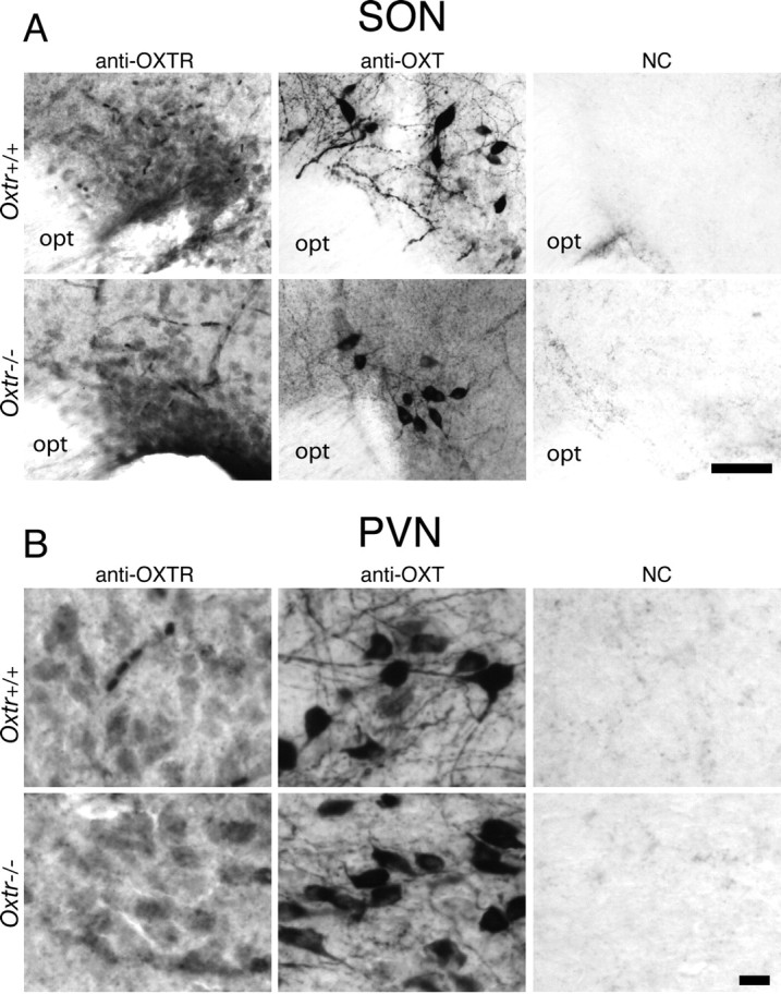
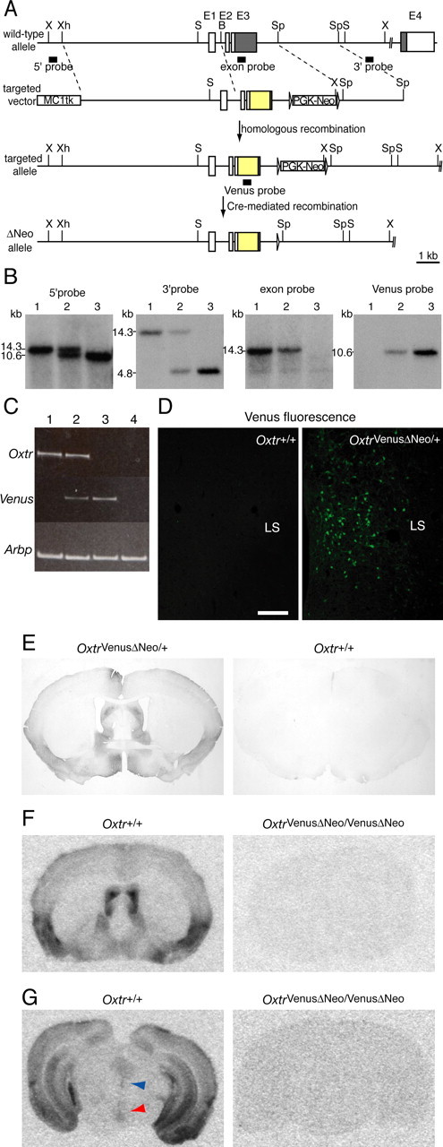
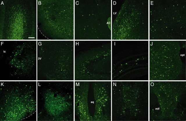
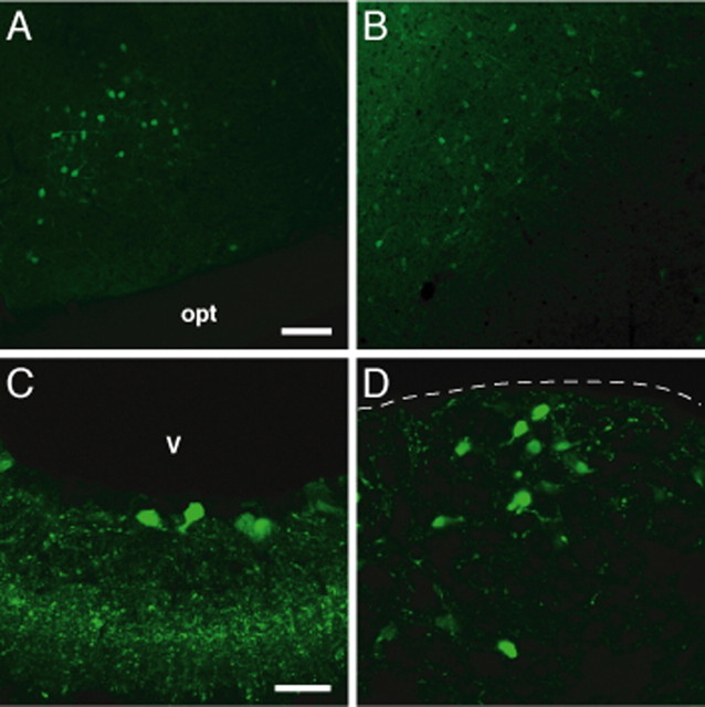
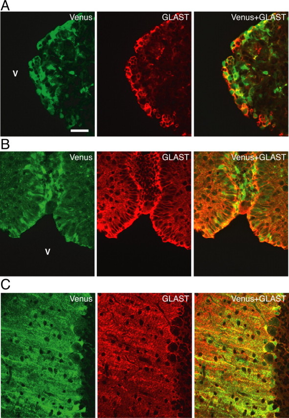
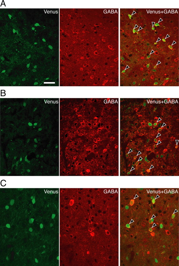
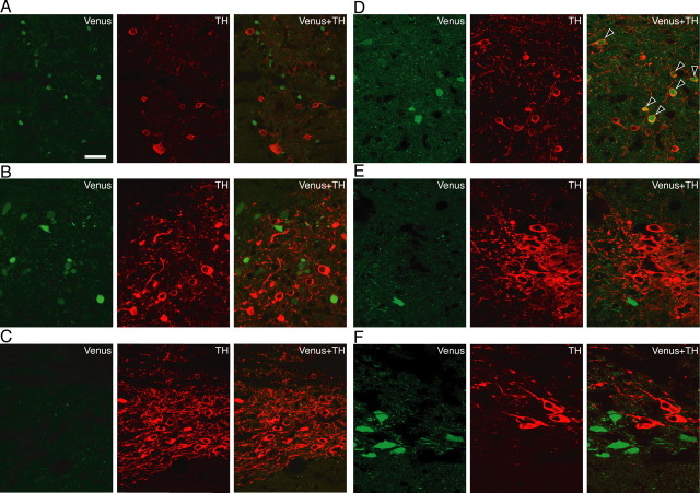
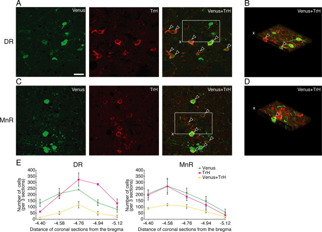
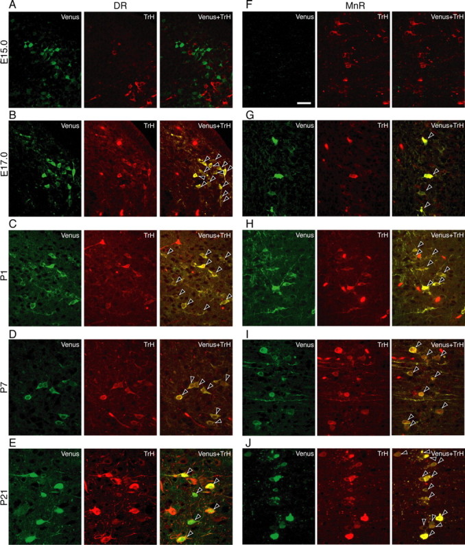
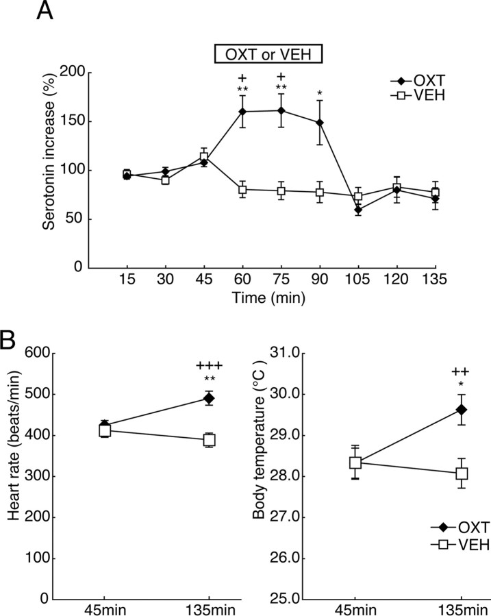
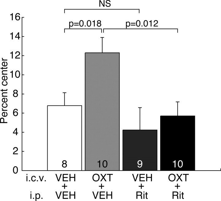
The oxytocin receptor has been implicated in the regulation of reproductive physiology as well as social and emotional behaviors. The neurochemical mechanisms by which oxytocin receptor modulates social and emotional behavior remains elusive, in part because of a lack of sensitive and selective antibodies for cellular localization. To more precisely characterize oxytocin receptor-expressing neurons within the brain, we generated an oxytocin receptor-reporter mouse in which part of the oxytocin receptor gene was replaced with Venus cDNA (a variant of yellow fluorescent protein). Examination of the Venus expression revealed that, in the raphe nuclei, about one-half of tryptophan hydroxylase-immunoreactive neurons were positive for Venus, suggesting a potential role for oxytocin in the modulation of serotonin release. Oxytocin infusion facilitated serotonin release within the median raphe nucleus and reduced anxiety-related behavior. Infusion of a 5-HT(2A/2C) receptor antagonist blocked the anxiolytic effect of oxytocin, suggesting that oxytocin receptor activation in serotonergic neurons mediates the anxiolytic effects of oxytocin. This is the first demonstration that oxytocin may regulate serotonin release and exert anxiolytic effects via direct activation of oxytocin receptor expressed in serotonergic neurons of the raphe nuclei. These results also have important implications for psychiatric disorders such as autism and depression in which both the oxytocin and serotonin systems have been implicated.


Figures

Figure 1.
Figure 1.
Immunohistochemistry using anti-OXTR and anti-OXT antibodies. In both Oxtr+/+ and Oxtr−/−, moderately immunoreactive cells for OXTR were found in SON (A) and PVN (B). The panels show immunoreactivity for OXTR (left), for OXT (middle), and negative control (NC) (no primary antibody) (right). Scale bars: SON, 50 μm; PVN, 10 μm. opt, Optic chiasm.
Figure 2.
Figure 2.
Generation of OXTR-Venus knock-in mice. A, The wild-type and mutant Oxtr loci and gene targeting constructs. Exons (E) are indicated by boxes (white boxes, 5′- and 3′-UTRs; gray boxes, coding regions). Venus-poly(A) is indicated by yellow boxes. Positions of restriction enzyme sites and the probes used for Southern blot analysis are shown (X, XbaI; Xh, XhoI; S, SacI; B, BamHI; Sp, SphI). The loxP sites are represented by arrowheads (not to scale). B, Southern blot analysis of genomic DNA from littermate progeny from intercrosses of OxtrVenus-Neo/+ mice. XbaI-digested tail DNA was hybridized with the radiolabeled probes indicated in Figure 1A. 5′ and 3′ probes external to the targeting vector and two internal probes were used to distinguish the wild-type (14.3 kb) and targeted alleles (10.6 kb for the 5′ and Venus probes, and 4.8 kb for the 3′ probe). Lane 1, Oxtr+/+; lane 2, OxtrVenus-Neo/+; lane 3, OxtrVenus-Neo/Venus-Neo. C, Oxtr and Venus mRNA expression in the mouse brain by reverse transcription (RT)-PCR. Total RNA extracted from the brains was subjected to RT-PCR. Lane 1, Oxtr+/+; lane 2, Oxtr VenusΔNeo/+; lane 3, OxtrVenusΔNeo/VenusΔNeo; lane 4, Oxtr−/−. D, Observation of Venus fluorescent in the LS of OxtrVenusΔNeo/+ mouse. E, Immunocytochemical detection of Venus protein using a DAB procedure in OxtrVenusΔNeo/+ and Oxtr+/+ mice. Venus immunoreactivity was observed as a brown precipitate. F, G, OXTR-binding autoradiograms in the brain of Oxtr+/+ (left) and OxtrVenusΔNeo/VenusΔNeo mice (right). OXTR binding was observed in the LS (F) and in the DR and MnR (G) of Oxtr+/+ mice, but not OxtrVenusΔNeo/VenusΔNeo mice. In G, blue and red arrowheads indicate the DR and MnR, respectively. Scale bar, 100 μm.
Figure 3.
Figure 3.
The distribution of Venus immunoreactivity in the brain of male OxtrVenusΔNeo/+ mice. Immunoreactive cells for Venus were found in GrO (A), Pir (B), AccC (C), LS (D), PFC (E), BST (F), MPA (G), CA2, CA3 (H), PoDG (I), MeA (J), CoA (K), CeA (L), PAG (M), MnR (N), and DR (O). Scale bar, 100 μm. AC, Anterior commissure; LV, lateral ventricle; ic, internal capsule; 3V, third ventricle; opt, optic chiasm; aq, aqueduct; mlf, medial longitudinal fasciculus.
Figure 4.
Figure 4.
The expression of Venus in the areas in which OXTR expression had not been described. Venus was detected by an immunocytochemical method with an anti-GFP antibody in LA (A), LH (B), ME (C), and AP (D). Scale bars: A, B, 100 μm; C, D, 30 μm. V, Ventricle.
Figure 5.
Figure 5.
Double immunocytochemical detection of Venus and GLAST, a marker of glial cells. GLAST-immunoreactive cells expressed Venus in the SFO (A), OVLT (B), and CCmol (C). Scale bar, 30 μm. V, Ventricle.
Figure 6.
Figure 6.
Colocalization of Venus with GABA. Venus and GABA were colocalized in the LS (A), BST (B), and CoA (C). The arrowheads indicate double-positive cells for Venus (green) and GABA (red). Scale bar, 30 μm.
Figure 7.
Figure 7.
Colocalization of Venus with TH. Areas of brain shown in each figure are as follows: Gl (A), Arc (B), VTA (C), PAG (D), LC (E), and NTS (F). TH-immunoreactive cells in the PAG were observed to express Venus. The arrowheads indicate double-positive cells for Venus (green) and TH (red). Scale bar, 30 μm.
Figure 8.
Figure 8.
Double labeling of Venus with TrH. Colocalization of Venus with TrH in the DR (A, B) and MnR (C, D). B and D show three-dimensional images constructed from serial tomographical images in open squares (A, C). E, Anteroposterior distribution of cells expressing immunoreactivity of Venus and/or TrH in the DR (left) and MnR (right) (n = 3). In the DR and MnR, about one-half of tryptophan hydroxylase-immunoreactive neurons were positive for Venus. Error bars indicate SEM. The arrowheads indicate double-positive cells for Venus (green) and TrH (red). Scale bar, 30 μm.
Figure 9.
Figure 9.
Developmental analysis of colocalization of Venus with TrH in the DR (A–E) and MnR (F–J). The brains of OxtrVenusΔNeo/+ mice were analyzed at various developmental ages of embryonic day 15 (E15) (A, F), E17 (B, G), and postnatal day 1 (P1) (C, H), P7 (D, I), and P21 (E, J). The arrowheads indicate double-positive cells for Venus (green) and TrH (red). Scale bar, 30 μm.
Figure 10.
Figure 10.
Facilitation of serotonin release in the MnR by local application of OXT. A, The average content in the first three perfusates of the MnR was set as 100%. One hundred percent for serotonin contents = 0.54 ± 0.10 (vehicle-perfused mice; n = 6) or 0.36 ± 0.05 pg/20 μl (OXT-perfused mice; n = 7). Serotonin release within the MnR was significantly facilitated during application of OXT but not the vehicle. *p < 0.05, **p < 0.01 compared with vehicle-perfused control group. +p < 0.05 compared with the first three perfusate samples. B, Effects of local application of OXT in the MnR on heart rates and body temperature. Heart rates (left) and body temperature (right) were significantly increased compared with those before OXT perfusion (45 min) or those of the vehicle-perfused control mice. *p < 0.05, **p < 0.01 compared with vehicle-perfused control group. ++p < 0.01, +++p < 0.001 compared with before OXT perfusion (45 min). Error bars indicate SEM.
Figure 11.
Figure 11.
Anxiolytic effect of intracerebroventricular administration of OXT and reversal by 5-HT2A/2C receptor antagonist. Intracerebroventricular administration of OXT (10 μg, i.c.v.) decreased anxiety-related behavior and the anxiolytic effect was blocked by coadministration of ritanserin (5 mg/kg, i.p.), a 5-HT2A/2C receptor antagonist in the open-field test. The number of mice in each group is indicated in the bottom of each bar. Total distance traveled in each group was 2903 ± 463, 6305 ± 622, 2819 ± 737, and 4197 ± 645 cm for mice injected with intracerebroventricular vehicle plus intraperitoneal vehicle, intracerebroventricular OXT plus intraperitoneal vehicle, intracerebroventricular vehicle plus intraperitoneal ritanserin, and intracerebroventricular OXT plus intraperitoneal ritanserin. Local application of OXT facilitated serotonin release within the raphe nucleus (Fig. 8A). Furthermore, anxiolytic effect of OXT was blocked by a 5-HT2A/2C receptor antagonist. Rit, Ritanserin; VEH, vehicle. Error bars indicate SEM.

Similar articles

Cited by

References

    1. Adan RA, Van Leeuwen FW, Sonnemans MA, Brouns M, Hoffman G, Verbalis JG, Burbach JP. Rat oxytocin receptor in brain, pituitary, mammary gland, and uterus: partial sequence and immunocytochemical localization. Endocrinology. 1995;136:4022–4028. - PubMed
    1. Aggleton JP. The amygdala: a functional analysis. Ed 2. London: Oxford UP; 2001.
    1. Bergquist F, Ludwig M. Dendritic transmitter release: a comparison of two model systems. J Neuroendocrinol. 2008;20:677–686. - PubMed
    1. Brunner D, Buhot MC, Hen R, Hofer M. Anxiety, motor activation, and maternal-infant interactions in 5HT1B knockout mice. Behav Neurosci. 1999;113:587–601. - PubMed
    1. Carter CS. Developmental consequences of oxytocin. Physiol Behav. 2003;79:383–397. - PubMed

Publication types

MeSH terms

LinkOut - more resources