. 2009 Feb 18;29(7):2297-308.
doi: 10.1523/JNEUROSCI.2034-08.2009.

Loss of the cholesterol-binding protein prominin-1/CD133 causes disk dysmorphogenesis and photoreceptor degeneration

Affiliations

Loss of the cholesterol-binding protein prominin-1/CD133 causes disk dysmorphogenesis and photoreceptor degeneration

Serena Zacchigna et al. J Neurosci. .

Abstract

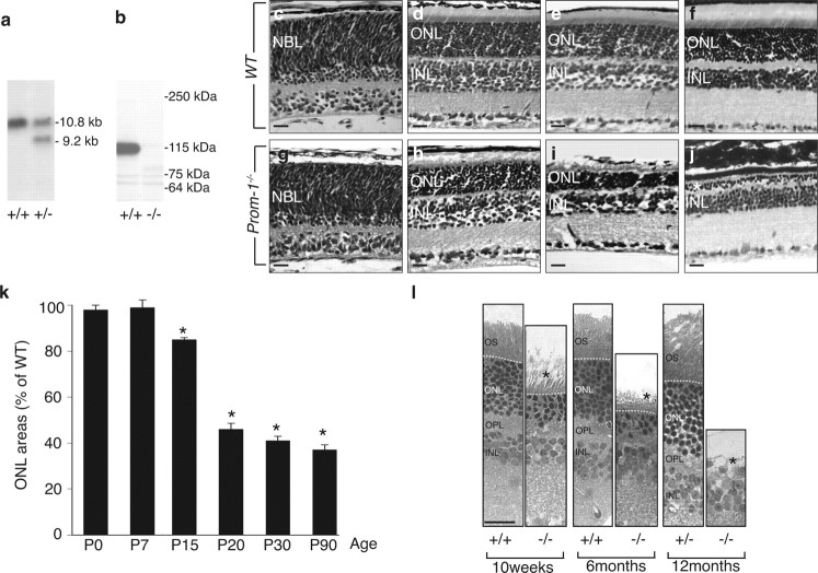
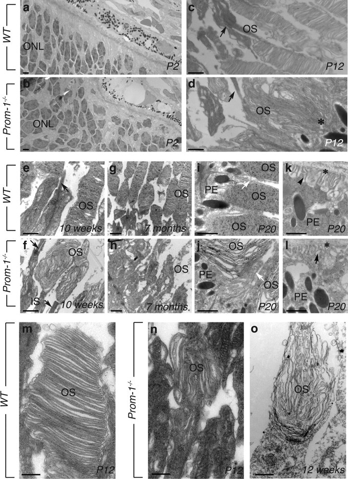
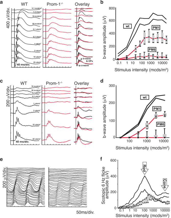
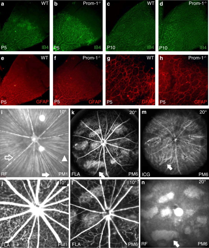
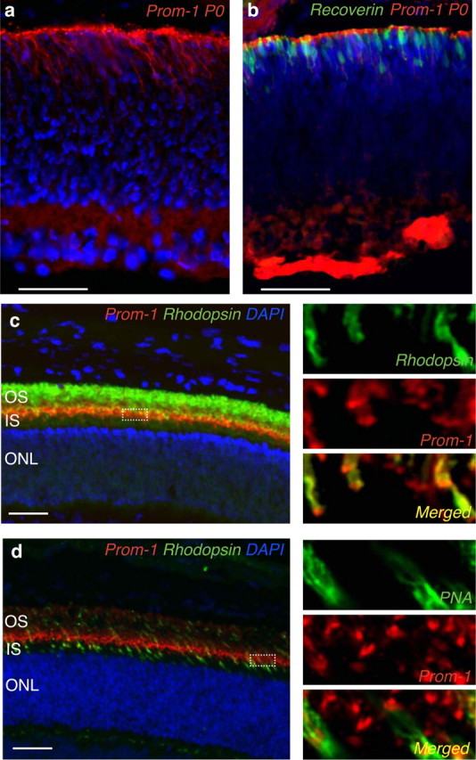
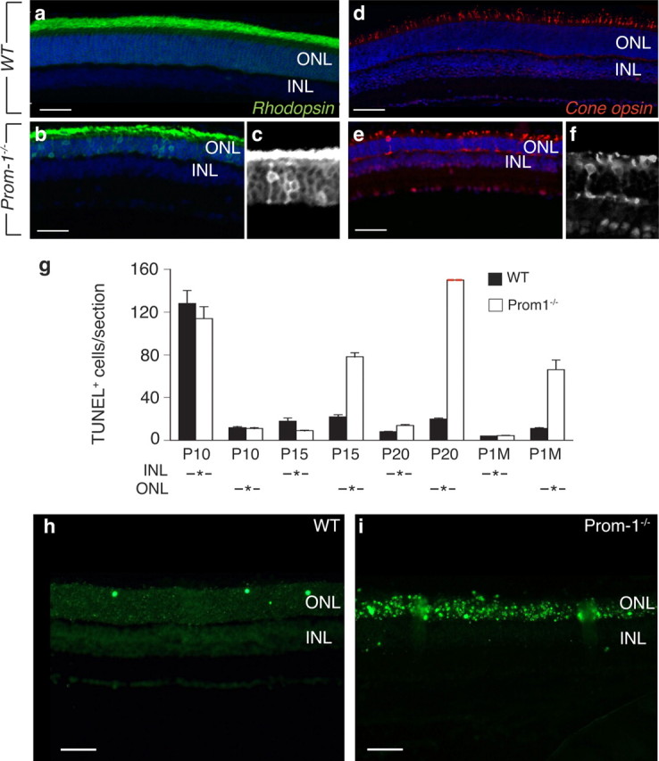
Prominin-1/CD133 (Prom-1) is a commonly used marker of neuronal, vascular, hematopoietic and other stem cells, yet little is known about its biological role and importance in vivo. Here, we show that loss of Prom-1 results in progressive degeneration of mature photoreceptors with complete loss of vision. Despite the expression of Prom-1 on endothelial progenitors, photoreceptor degeneration was not attributable to retinal vessel defects, but caused by intrinsic photoreceptor defects in disk formation, outer segment morphogenesis, and associated with visual pigment sorting and phototransduction abnormalities. These findings shed novel insight on how Prom-1 regulates neural retinal development and phototransduction in vertebrates.


Figures

Figure 1.
Figure 1.
Absence of Prom-1 leads to retina degeneration. a, Southern blot of BamHI-NciI-digested genomic DNA from ES cell clones generated a unique 10.8 kb band in WT (left lane) and an additional 9.2 kb allele in Prom-1+/− clones (right lane). b, Western blot analysis with an antibody (13A4) for Prom-1 on extracts from WT (left lane) and Prom-1−/− (right lane) kidney confirmed the deletion of Prom-1 protein (115 kDa) in the knock-out mouse. c–k, A time course morphometric analysis of ONL areas clearly indicates that the ONL (NBL for P0) area progressively decreases in Prom-1−/− mice. Shown are hematoxylin and eosin staining of WT (c–f) and Prom-1−/− (g–j) retinal sections at P0 (c, g), P15 (d, h), P20 (e, i), and 6 months (f, j). The quantification of the ONL area over time confirmed that degeneration becomes morphologically evident in Prom-1−/− mice starting from P15 (k). *p < 0.05 versus WT. l, Semithin sections (1 μm) of 10-week-, 6-month- and 12-month-old mice stained with toluidine blue showing that the thickness of the ONL is severely reduced in KO mice at 10 and 24 weeks of age and completely missing in older Prom-1−/− animals. No significant differences are evident in Prom-1+/− mice. OPL, Outer plexiform layer. Dashed line, external limiting membrane. The asterisk in j and l indicates the degenerated photoreceptor layer. Scale bar, 20 μm.
Figure 2.
Figure 2.
Abnormal OS morphogenesis in Prom-1−/− mice. Time course study of OS morphology by TEM. a, b, Ultrastructure analyses of WT (a) and Prom-1−/− (b) retinas at P2 shows that in both cases the OS is not formed yet. c, d, At P12, the OS of Prom-1−/− mice appears already abnormal (black asterisk in d), indicating that the OS membranes are misfolded early during their development. At later stages, a progressive disorganization of the rod OS is evident in Prom-1−/− mice at 10 weeks (compare f with e) and 7 months (compare h with g), when the photoreceptors have almost disappeared. Black arrows in cf indicate connecting cilia. In contrast, the RPE layer is well preserved in Prom-1−/− retina, with intact microvilli (white arrows in i and j) and basal infoldings (black arrowheads in k and l; asterisk, Bruch' membrane). m–o, Ultrastracture analysis of cones, as recognized by their position and PNA labeling, followed by ultrasmall gold detection and silver enhancement. Whereas the disks appear regularly stacked in the OS of cones in WT mice at P12 (m), they are completely disarranged in Prom-1−/− retinas (n), indicating that both rods and cones already display abnormal OS structures at this age. Even more profound alterations are evident in PNA-labeled cones at 12 weeks (o). Black dots are silver particles representing the PNA staining of the OS and IS of the cone cells. IS, Inner segment; PE, pigmented epithelial cells. Scale bars: a–l, 1 μm, m–o, 200 nm.
Figure 3.
Figure 3.
Impaired rod and cone function in Prom-1−/− mice. Functional evaluation of Prom-1−/− mice at 1 month (PM1) and 6 months (PM6) by Ganzfeld ERG. a, b, Scotopic (dark-adapted) single-flash recordings. a, Comparison of representative records between Prom-1−/− mice and WT controls at PM1. b, Statistical evaluation (box-and-whisker plot) of Prom-1−/− mice at PM1 and PM6 compared with WT mice (PM1). The two black lines indicate the normal range (5 to 95% quantile). The red lines give the median, the gray boxes the 25 and 75% quantiles, and the black whiskers the 5 and 95% quantiles of Prom-1−/− mice at PM1 and PM6, respectively. c, d, Photopic (light-adapted) single-flash recordings. c, Comparison of representative records between Prom-1−/− mice and WT controls at PM1. d, Statistical evaluation (box-and-whisker plot as above) of Prom-1−/− mice at PM1 and PM6 compared with WT mice (PM1). e, f, Scotopic flicker recordings at 6Hz flash frequency. e, Comparison of representative records between Prom-1−/− mice (right) and WT controls (left) at PM1. f, Statistical evaluation (box-and-whisker plot) of Prom-1−/− mice compared with WT mice at PM1. The right shift of both rod and cone peaks indicates a reduction in sensitivity to light, and the drop in amplitude an additional reduction of functional cell numbers.
Figure 4.
Figure 4.
Development and involution of the vascular system in Prom-1−/− retinas. a–h, Normal development of retinal vessels and astrocytic templates in Prom-1−/− retinas. Whole mount staining for isolectin IB4 showing a comparable development of retinal vasculature at P5 in Prom-1−/− retinas (b) and WT mice (a). An equivalent pattern of Isolectin IB4 staining was also observed in WT (c) and Prom-1−/− (d) retinas at P10. GFAP staining at P5 reveals that astrocytic networks also develop similarly in both WT (e) and Prom-1−/− retinas (f). A higher magnification view of peripheral retinas shows that GFAP-positive networks reach fully to the periphery in both WT (g) and Prom-1−/− retinas (h). i–n, Involution of retinal vessels during the course of degeneration in Prom-1−/− mice. Retinal arteries (open arrow) and veins (filled arrow) in Prom-1−/− retinas appeared normal at 1 month postnatally (PM1), as shown by red-free (RF) surface imaging (514 nm, 10°; i), and fluorescence angiography (FLA, 488 nm, barrier at 500 nm, 10°; j). k–n, At 6 months postnatally (PM6), the vasculature appears strongly attenuated. FLA (20°) shows reduction of retinal capillaries, reduced diameter and caliber fluctuations of large retinal vessels, and extended areas of choriocapillaris/RPE loss (arrow) (k). l, Magnification of (k), i.e., FLA (10°), same area as in j. ICG angiography (795 nm, barrier at 800 nm, 20°) enhances the view of choroidal structures because of less absorption by melanin (m). The arrow points to the same area as in (k). An RF surface image (20°) most clearly depicts the lesions (n). The arrow points to the same area as in k and m.
Figure 5.
Figure 5.
Localization of Prom-1 protein in WT retina. a, b, Immunofluorescent staining (red) shows that at P0 Prom-1 protein is expressed at the interface between the neural and epithelial retina (a), when developing photoreceptors can be recognized by recoverin staining (b). c, d, Confocal microscopy showing Prom-1 expression at P20 at the basal part of the OS of adult rods and cones, as assessed by double immunostaining for Prom-1 in red and either rhodopsin (c) or PNA (d) in green. The higher magnification insets on the right part of each figure better highlight the subcellular localization of Prom-1 in rods and cones, as shown by the yellow color resulting in the merged pictures (yellow, lower insets). IS, Inner segment. Scale bar, 50 μm.
Figure 6.
Figure 6.
Abnormal pigment sorting and photoreceptor apoptosis in Prom-1−/− mice. a–f, Pigment mislocalization in Prom-1−/− retinas. Rhodopsin immunostaining reveals pigment mislocalization at P20 in Prom-1−/− retinas (b), where the ONL still retains rhodopsin signal, while almost no staining is evident in ONL of WT retinas (a). Similarly, cone opsin staining at P20 reveals shortened OS in cone photoreceptors of Prom-1−/− retinas and pigment mis-sorting to the ONL and the synaptic space (e) compared with WT retinas (d). Black and white magnification insets (c, f) better highlight the abnormal localization of rod and cone opsin, respectively. g, A time course study of TUNEL assay shows an increased number of apoptotic cells in the ONL starting at P15 in Prom-1−/− retinas, with a dramatic peak at P20, when the number of apoptotic cells was not countable (red dashed line). Data are presented as the mean number of TUNEL+ cells per section in the INL or ONL area. h, i, Representative pictures of TUNEL staining, showing the presence of massive apoptosis in Prom-1−/− (i) ONL, compared with WT (h) at P20. Scale bars: a–f, 100 μm; h, i, 50 μm.

References

    1. Adamian M, Pawlyk BS, Hong DH, Berson EL. Rod and cone opsin mislocalization in an autopsy eye from a carrier of X-linked retinitis pigmentosa with a Gly436Asp mutation in the RPGR gene. Am J Ophthalmol. 2006;142:515–518. - PubMed
    1. Bascom RA, Manara S, Collins L, Molday RS, Kalnins VI, McInnes RR. Cloning of the cDNA for a novel photoreceptor membrane protein (rom-1) identifies a disk rim protein family implicated in human retinopathies. Neuron. 1992;8:1171–1184. - PubMed
    1. Carmeliet P, Tessier-Lavigne M. Common mechanisms of nerve and blood vessel wiring. Nature. 2005;436:193–200. - PubMed
    1. Corbeil D, Röper K, Fargeas CA, Joester A, Huttner WB. Prominin: a story of cholesterol, plasma membrane protrusions and human pathology. Traffic. 2001;2:82–91. - PubMed
    1. Deretic D. A role for rhodopsin in a signal transduction cascade that regulates membrane trafficking and photoreceptor polarity. Vision Res. 2006;46:4427–4433. - PubMed

Publication types

MeSH terms