. 2009 Mar;119(3):650-60.
doi: 10.1172/JCI37617. Epub 2009 Feb 23.

Parkin, PINK1, and DJ-1 form a ubiquitin E3 ligase complex promoting unfolded protein degradation

Affiliations

Parkin, PINK1, and DJ-1 form a ubiquitin E3 ligase complex promoting unfolded protein degradation

Hui Xiong et al. J Clin Invest. 2009 Mar.

Abstract

Mutations in PARKIN, pten-induced putative kinase 1 (PINK1), and DJ-1 are individually linked to autosomal recessive early-onset familial forms of Parkinson disease (PD). Although mutations in these genes lead to the same disease state, the functional relationships between them and how their respective disease-associated mutations cause PD are largely unknown. Here, we show that Parkin, PINK1, and DJ-1 formed a complex (termed PPD complex) to promote ubiquitination and degradation of Parkin substrates, including Parkin itself and Synphilin-1 in neuroblastoma cells and human brain lysates. Genetic ablation of either Pink1 or Dj-1 resulted in reduced ubiquitination of endogenous Parkin as well as decreased degradation and increased accumulation of aberrantly expressed Parkin substrates. Expression of PINK1 enhanced Parkin-mediated degradation of heat shock-induced misfolded protein. In contrast, PD-pathogenic Parkin and PINK1 mutations showed reduced ability to promote degradation of Parkin substrates. This study identified a functional ubiquitin E3 ligase complex consisting of PD-associated Parkin, PINK1, and DJ-1 to promote degradation of un-/misfolded proteins and suggests that their PD-pathogenic mutations impair E3 ligase activity of the complex, which may constitute a mechanism underlying PD pathogenesis.


Figures

Figure 1
Figure 1. Complex formation of Parkin, PINK1, and DJ-1.
(AF) Association of Parkin, PINK1, and DJ-1 in transfected cells. Parkin-VSVG, PINK1-flag, and DJ-1–myc were expressed in various combinations and immunoprecipitated with antibodies to the corresponding tag, followed by detection of coprecipitation of PINK1 and DJ-1 (A), Parkin and DJ-1 (B), and Parkin and PINK1 (C), respectively. (DF) Inputs of Parkin (D), PINK1 (E), and DJ-1 (F). Note that cotransfection of PINK1 significantly reduced Parkin levels in lysates. Tub, cytosolic marker tubulin. (G and H) In vitro assembly of the PPD complex. Affinity-purified Parkin-myc-flag (Parkin), PINK1-VSVG-flag (PINK1), and GST–DJ-1–VSVG (DJ-1GST) were incubated in various combinations, followed by precipitation with either anti-myc agarose (G) or GST agarose (H). Precipitates were detected with an anti-VSVG antibody to detect both PINK1 and DJ-1–GST (G), an anti-Parkin antibody to detect Parkin (H), an anti-PINK1 antibody to detect PINK1 (H), or an anti–DJ-1 antibody to detect DJ-1 (H). (I) Association of Parkin, PINK1, and DJ-1 in vivo. Lysates of human brain cortex from 2 unrelated individuals (lanes 1 and 2 for one individual, lanes 3 and 4 for the other) were immunoprecipitated with an anti-Parkin monoclonal antibody (αParkin) or control mouse IgG (mIgG), followed by immunoblotting with a polyclonal anti-Parkin antibody, a polyclonal anti-PINK1 antibody, or a monoclonal anti–DJ-1 antibody. Multiple endogenous PINK1 proteolytic bands were detected (arrows). (J) A schematic illustration of interaction among PPD complex components. IBR, in between RING fingers; MTS, mitochondrial targeting sequence; UBL, ubiquitin-like.
Figure 2
Figure 2. Detection of the PPD complex in both mitochondria and cytosolic fractions.
Cells expressing Parkin alone or a combination of Parkin, PINK1, and DJ-1 were fractionated to mitochondrial (Mito) and cytosolic (Cyto) fractions. Left: Expression of Parkin, PINK1, DJ-1, mitochondria marker complex I (Cplx I), and cytosolic marker tubulin. Right: Coimmunoprecipitation of Parkin with PINK1 and DJ-1 in mitochondria and cytosolic fractions.
Figure 3
Figure 3. PINK1 promotes proteasomal degradation of Parkin and Synphilin-1.
(A and B) SH-SY5Y cells coexpressing PINK1-flag and Parkin-VSVG (A) or PINK1-flag and Synphilin-1–EGFP (Syn-1; B) were treated with either MG132 or leupeptin (Leu). Steady-state levels of Parkin, Synphilin-1, PINK1, and actin (loading control) are shown. (C and D) Expression of PINK1 reduced Parkin stability via the proteasomal pathway. Cells transfected with Parkin alone (C; top panel), Parkin and PINK1 (C; middle panel), or Parkin and PINK1 with MG132 treatment (C; bottom panel) were pulse-labeled, followed by chasing for the indicated time intervals. Levels of Parkin were detected by immunoprecipitation. Results from a representative experiment (C) and quantitation of 3 independent experiments are shown (D). Multiple Parkin bands likely representing ubiquitinated Parkin were detected in the presence of PINK1 (arrows). (D) Diamonds indicate Parkin alone, squares indicate Parkin and PINK1, and triangles indicate Parkin and PINK1 with MG132 treatment.
Figure 4
Figure 4. PINK1 regulates ubiquitination of Parkin and Synphilin-1.
Parkin (left) or Synphilin-1 (right) was cotransfected into SH-SY5Y cells with PINK1WT, a PD-pathogenic PINK1G309D mutant (PINK1m), DJ-1WT, or a PD-pathogenic DJ-1L166P mutant (DJ-1m) in the presence of ubiquitin (Ub) in various combinations. In a Synphilin-1 degradation experiment, a PD-pathogenic ParkinR42P mutant (Parkinm) was included. Parkin (A and B) or Synphilin-1 (G and H) were immunoprecipitated from equal amounts of cell lysates, followed by detection of ubiquitin (A and G), Parkin (B), or Synphilin-1 (H). Expression levels of DJ-1WT (C), DJ-1L166P (C), and ubiquitin (D and J) were shown by direct immunoblotting. Ubiquitination and steady-state levels of PINK1 variants were observed by immunoprecipitation of PINK1, followed by immunoblotting of either ubiquitin (E and K) or PINK1 (F and L).
Figure 5
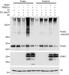
Figure 5. PINK1 promotes Parkin auto-ubiquitination in vitro.
Affinity-purified ParkinWT, the PD-associated ParkinR42P mutant, PINK1WT, and PD-associated PINK1G309D mutant proteins in various combinations were assayed for in vitro ubiquitination in the presence of recombinant E1, E2 (Ubc7), and HA-tagged ubiquitin. Proteins were separated on SDS-PAGE and immunoblotted with an anti-Parkin antibody to detect Parkin monomers (Parkin) and ubiquitinated Parkin [Parkin-poly(Ub)], an anti-VSVG antibody to detect PINK1, or an anti-HA antibody to detect ubiquitin. Ubiquitinated Parkin appeared as a smear in the top panel.
Figure 6
Figure 6. Ubiquitination of endogenous Parkin in mouse brains with PINK1 and DJ-1 ablation.
Top: Brain slices from WT and PINK1-deficient (KO) mice were immunoprecipitated with either a monoclonal anti-Parkin antibody or a control mouse IgG, followed by immunoblotting with an anti-ubiquitin antibody. Cells overexpressing exogenous Parkin were used as a positive control. The experiments were done with or without heat shock treatment (HS). Bottom: Immunoprecipitated Parkin protein was detected by anti-Parkin polyclonal antibody. The 55-kDa band shown in control precipitations were IgG heavy chain. Note that Parkin ubiquitination was remarkably reduced in PINK1-null brain slices.
Figure 7
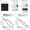
Figure 7. Genetic ablation of mouse Pink1 or Dj-1 results in increased stability of aberrantly expressed Parkin.
(A) Expression of Parkin, PINK1, and DJ-1 in PINK1- or DJ-1–deficient mouse fibroblasts. RT-PCR detection of Parkin, PINK1, and DJ-1 in PINK1 WT, PINK1 KO, DJ-1 WT, and DJ-1 KO cells. Control, no cDNA template added. (B and C) Increased accumulation of aberrantly expressed Parkin in PINK1 KO and DJ-1 KO cells. Cells transfected with control plasmid or plasmid encoding Parkin showed increased Parkin detection in PINK1 KO cells (B) and DJ-1 KO cells (C). (C) Tubulin was used as a control. Lack of DJ-1 protein in DJ-1 KO cells was shown by immunoblotting. (DG) Increased stability of Parkin in PINK1 KO and DJ-1 KO cells. PINK1 KO, PINK1 WT, DJ-1 KO, and DJ-1 WT cells were transfected with Parkin, followed pulse chase analysis of Parkin stability for the time frames indicated. Representative results of PINK1 (D) and DJ-1 (E) are shown. Quantitation was obtained from PINK1 KO cells generated from 2 independent PINK1 KO mice (F) and DJ-1 KO cells generated from multiple DJ-1 KO mice (G).
Figure 8
Figure 8. PINK1 promotes degradation of Parkin that has accumulated as a result of heat shock treatment.
Cells expressing Parkin alone (lanes 2–4); Parkin and ubiquitin (lanes 5–7); and Parkin, ubiquitin, and PINK1 (lanes 8–10) without heat shock treatment (lanes 1, 2, 5, 8), with heat shock treatment (lanes 3, 6, 9), or with heat shock treatment followed by a 4-h recovery time (RT) at 37°C (lanes 4, 7, 10). The cells were lysed and immunoprecipitated with an anti-Parkin antibody, followed by immunoblotting with either an anti-ubiquitin antibody (A) or an anti-Parkin antibody (B). Expression of PINK1 (C) and tubulin (D) were shown with immunoblotting. Note that heat shock treatment increased accumulation of Parkin protein (B; lanes 6–7). PINK1 promoted degradation of Parkin even with heat shock treatment (B; lanes 9–10).
Figure 9
Figure 9. PD-pathogenic Parkin or PINK1 mutants impair Parkin degradation.
(A) Interaction of PINK1WT with PD-pathogenic ParkinΔE4. Cells expressing PINK1WT alone (Control), PINK1WT and ParkinΔE4 (ΔE4), or PINK1WT and ParkinWT (Parkin) were immunoprecipitated with an anti-flag antibody (to precipitate PINK1), followed by immunoblotting with an anti-myc antibody (to detect Parkin variants). (B) Interaction of ParkinWT with PINK1G309D. Cells expressing ParkinWT alone (control), ParkinWT and PINK1G309D (G309D), or ParkinWT and PINK1WT (PINK1) were immunoprecipitated with anti-myc antibody (to precipitate Parkin), followed by immunoblotting with an anti-flag antibody (to detect PINK1 variants). (C) PINK1 promoted degradation of PD-pathogenic Parkin mutants. ParkinWT (WT) and PD-pathogenic ParkinR42P, -T240W, and -ΔE4 were cotransfected with or without PINK1. Steady-state levels of Parkin, PINK1, and tubulin were analyzed by immunoblotting. PINK1 promoted significant degradation of ParkinWT but not ParkinR42P, -T240W, or -ΔE4. (D) PD-pathogenic PINK1 mutants were impaired in promoting Parkin degradation. PINK1WT (WT) and PD-pathogenic PINK1G309D, -T313M, and -P399L were cotransfected with or without Parkin. Steady-state levels of Parkin, PINK1, and tubulin were detected by immunoblotting. PD-pathogenic mutants showed little or reduced ability to promote Parkin degradation. (E and F) Quantitation of Parkin degradation affected by pathogenic Parkin (E) or PINK1 (F) mutations. The data were from 3 independent experiments. Relative Parkin levels were normalized to either the level of Parkin variants without PINK1 expression (E, black bars) or the level of ParkinWT without PINK1 expression (Ctrl; F) in the same experiment.

Comment in

Similar articles

Cited by

References

    1. Dawson T.M., Dawson V.L. Molecular pathways of neurodegeneration in Parkinson’s disease. Science. 2003;302:819–822. doi: 10.1126/science.1087753. - DOI - PubMed
    1. Valente E.M., et al. Hereditary early-onset Parkinson’s disease caused by mutations in PINK1. Science. 2004;304:1158–1160. doi: 10.1126/science.1096284. - DOI - PubMed
    1. Paisan-Ruiz C., et al. Cloning of the gene containing mutations that cause PARK8-linked Parkinson’s disease. Neuron. 2004;44:595–600. doi: 10.1016/j.neuron.2004.10.023. - DOI - PubMed
    1. Zimprich A., et al. Mutations in LRRK2 cause autosomal-dominant parkinsonism with pleomorphic pathology. Neuron. 2004;44:601–607. doi: 10.1016/j.neuron.2004.11.005. - DOI - PubMed
    1. Cookson M.R. The biochemistry of Parkinson’s disease. Annu. Rev. Biochem. 2005;74:29–52. doi: 10.1146/annurev.biochem.74.082803.133400. - DOI - PubMed

Publication types

MeSH terms