. 2009 Mar;119(3):454-64.
doi: 10.1172/JCI37613. Epub 2009 Feb 23.

Combinatorial drug design targeting multiple cancer signaling networks controlled by mitochondrial Hsp90

Affiliations

Combinatorial drug design targeting multiple cancer signaling networks controlled by mitochondrial Hsp90

Byoung Heon Kang et al. J Clin Invest. 2009 Mar.

Abstract

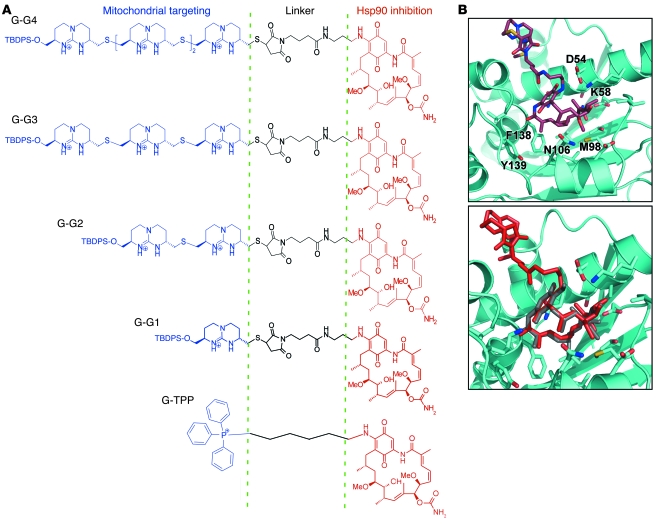
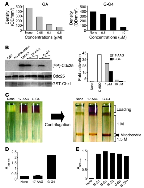
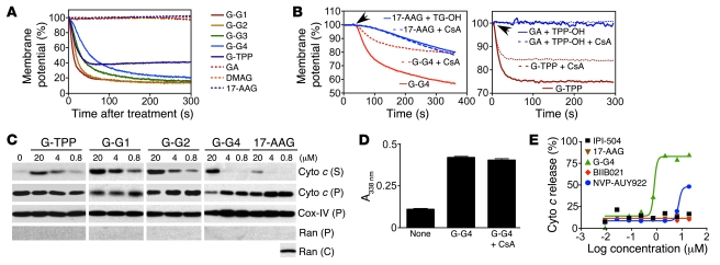
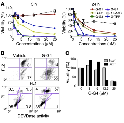
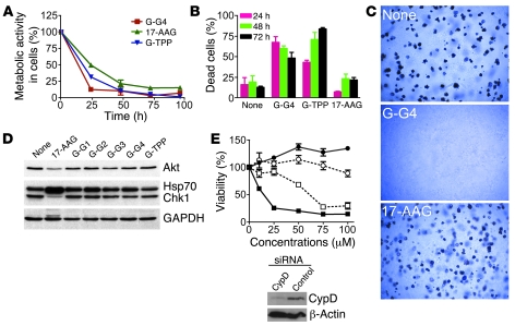
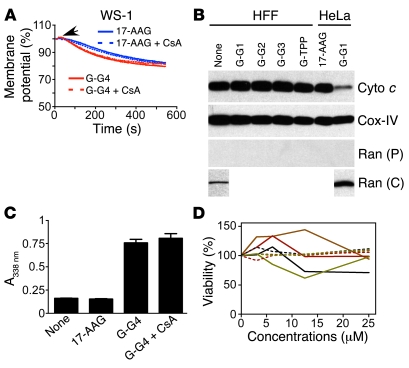
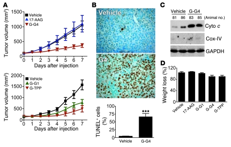
Although therapeutically targeting a single signaling pathway that drives tumor development and/or progression has been effective for a number of cancers, in many cases this approach has not been successful. Targeting networks of signaling pathways, instead of isolated pathways, may overcome this problem, which is probably due to the extreme heterogeneity of human tumors. However, the possibility that such networks may be spatially arranged in specialized subcellular compartments is not often considered in pathway-oriented drug discovery and may influence the design of new agents. Hsp90 is a chaperone protein that controls the folding of proteins in multiple signaling networks that drive tumor development and progression. Here, we report the synthesis and properties of Gamitrinibs, a class of small molecules designed to selectively target Hsp90 in human tumor mitochondria. Gamitrinibs were shown to accumulate in the mitochondria of human tumor cell lines and to inhibit Hsp90 activity by acting as ATPase antagonists. Unlike Hsp90 antagonists not targeted to mitochondria, Gamitrinibs exhibited a "mitochondriotoxic" mechanism of action, causing rapid tumor cell death and inhibiting the growth of xenografted human tumor cell lines in mice. Importantly, Gamitrinibs were not toxic to normal cells or tissues and did not affect Hsp90 homeostasis in cellular compartments other than mitochondria. Therefore, combinatorial drug design, whereby inhibitors of signaling networks are targeted to specific subcellular compartments, may generate effective anticancer drugs with novel mechanisms of action.


Figures

Figure 1
Figure 1. Structure of Gamitrinibs.
(A) Combinatorial modular design. The indicated individual modules of Gamitrinibs (mitochondrial targeting, linker, and Hsp90 inhibition) are indicated. G-G1, Gamitrinib-G1; G-TPP, Gamitrinib–TPP-OH; TBDPS, tert-butyldiphenylsilyl. (B) A 3D docking model of Gamitrinib-G1 with Hsp90 N-domain (top panel). The side chains of contact sites are labeled and in color. An overlay of Gamitrinib-G1 (red) and GA (gray) in the ATPase pocket of Hsp90 (bottom panel).
Figure 2
Figure 2. Inhibition of Hsp90 chaperone activity.
(A) Competition with GA affinity beads. GA (left panel) or Gamitrinib-G4 (right panel), at the indicated concentrations, were incubated with aliquots of SKBr3 tumor cell lysates followed by affinity purification of Hsp90 using GA affinity beads. Data are densitometric quantifications of scanning and image analysis of Hsp90 bands visualized by Western blotting. (B) Inhibition of Chk1 kinase activity. 17-AAG or Gamitrinib-G4 (1–10 μM) were analyzed for modulation of Chk1-dependent phosphorylation of Cdc25. Densitometric quantification of protein bands (right panel). (A and B) Data are representative of 2 independent experiments with identical results. (C) Accumulation of Gamitrinib in mitochondria. Cellular extracts were loaded with vehicle (None), unconjugated 17-AAG, or Gamitrinib-G4 and analyzed before (left panel) or after gradient density ultracentrifugation (right panel). The position of the 1–1.5 M interface corresponding to isolated mitochondria is indicated. (D) Quantification of mitochondrial accumulation. Mitochondria isolated from HeLa cells were incubated with 17-AAG, Gamitrinib-G4, or vehicle and analyzed using absorbance. Data are the mean ± SEM (n = 3). (E) Mitochondriotropic properties of all Gamitrinibs. Mitochondria isolated from Raji cells were incubated with the indicated Gamitrinibs or vehicle and analyzed by absorbance. Data are from a representative experiment out of 2 independent determinations.
Figure 3
Figure 3. Mitochondrial dysfunction.
(A) Mitochondrial membrane depolarization. Tetramethylrhodamine methyl ester–loaded (TMRM-loaded) HeLa cell mitochondria treated with Gamitrinibs or the various indicated agents (1 μM) were analyzed for changes in fluorescence emission. (B) Analysis of individual mitochondriotropic moieties. TMRM-loaded HeLa cell mitochondria were incubated with 1.5 μM 17-AAG plus tetraguanidinium (TG-OH) or Gamitrinib-G4 (left panel) or 0.7 μM GA plus TPP-OH or Gamitrinib–TPP-OH (right panel) and analyzed for changes in fluorescence emission in the presence or absence of CsA (5 μM). Arrows indicate point of addition. (C) Cytochrome c (Cyto c) release. Mitochondria isolated from HeLa cells treated with Gamitrinibs or 17-AAG (20 minutes) were analyzed for cytochrome c release in supernatants (S) or pellets (P). Cox-IV or Ran was used as a mitochondrial or cytosolic marker, respectively. Reactivity of the antibody to Ran with isolated cytosolic extracts (C) was used as a control. (D) Mitochondrial accumulation. Isolated HeLa cell mitochondria were incubated with vehicle or Gamitrinib-G4 in the presence or absence of CsA and analyzed using absorbance. Data are the mean ± SEM. (E) Analysis of nontargeted Hsp90 inhibitors. Isolated HeLa cell mitochondria were incubated with increasing concentrations of the various indicated agents for 3 hours and analyzed for cytochrome c release. Data are representative of 2 independent experiments.
Figure 4
Figure 4. Gamitrinib-mediated anticancer activity.
(A) Time course. H460 cells treated with the indicated concentrations of Gamitrinibs or 17-AAG were analyzed by a 3-(4,5-dimethylthiazol-2-yl)-2,5-diphenyltetrazolium bromide (MTT) assay after 3 hours (left panel) or 24 hours (right panel). Data are the mean ± SD (n = 2). (B) Caspase-associated cell death. H460 cells, treated with Gamitrinib-G4 or vehicle for 4 hours, were labeled with JC-1 and analyzed for loss of mitochondrial membrane potential by changes in FL2/FL1 fluorescence ratio (top panel) or Asp-Glu-Val-Asp-ase (DEVDase) (caspase) activity (bottom panel) using multiparametric flow cytometry. The percentage of cells in each quadrant is indicated. PI, propidium iodide. (C) Insensitivity to Bax. Wild-type or Bax–/– HCT116 cells were incubated with the indicated increasing concentrations of Gamitrinib-G4 and analyzed after 6 hours using MTT assay. Data are representative of 2 experiments.
Figure 5
Figure 5. Gamitrinibs induction of mitochondrial apoptosis.
(A) Comparison with 17-AAG. SKBr3 cells were treated with Gamitrinibs or 17-AAG (10 μM) for the indicated time intervals and analyzed using MTT assay. Data are representative of at least 2 independent experiments. (B) Tumor cell killing. SKBr3 cells treated with vehicle, Gamitrinib-G4, Gamitrinib–TPP-OH, or 17-AAG (10 μM), for the indicated time intervals, were analyzed by Trypan blue exclusion. Data are the mean ± SEM (n = 3). (C) Colony formation. H460 cells treated with vehicle, 17-AAG (50 μM), or Gamitrinib-G4 (50 μM) for 4 hours were analyzed for colony formation in soft agar after 2 weeks. Representative microscopy fields are shown. Original magnification, ×40. (D) Client protein modulation. HeLa cells treated with the indicated Gamitrinibs, 17-AAG (5 μM), or vehicle were analyzed for modulation of Hsp90 client proteins Akt and Chk1 in the cytosol or changes in expression of Hsp70 after 24 hours by Western blotting. (E) Requirement for CypD in Gamitrinib anticancer activity. H460 cells transfected with control (closed symbols) or CypD (open symbols) siRNA were treated with 17-AAG (circles) or Gamitrinib-G4 (squares) and analyzed using MTT assay after 6 hours. Data are the mean ± SEM (n = 3). The inset shows Western blotting of CypD knockdown by siRNA.
Figure 6
Figure 6. Selectivity of Gamitrinib anticancer activity.
(A) Mitochondrial membrane potential. TMRM-loaded mitochondria isolated from WS-1 normal human fibroblasts were incubated with Gamitrinib-G4 or 17-AAG plus the uncoupled mitochondriotropic moiety TG-OH and analyzed for changes in inner membrane potential in the presence or absence of CsA. (B) Cytochrome c release. Mitochondria isolated from normal HFF fibroblasts were treated with Gamitrinibs or 17-AAG and analyzed by Western blotting. HeLa cells were used as control. Cox-IV or Ran was used as a mitochondrial or cytosolic marker, respectively. Reactivity of the antibody to Ran with isolated cytosolic extracts from HeLa or HFF cells was used as a control. (C) Mitochondrial accumulation. Isolated normal mouse liver mitochondria were incubated with vehicle, 17-AAG, or Gamitrinib-G4 in the presence or absence of CsA and analyzed using absorbance. Data are the mean ± SEM. (D) Analysis of cell viability. Human fibroblasts (HFF, black line), bovine aortic endothelial cells (brown line), intestinal epithelial cells (red line), or human umbilical vein endothelial cells (green line) were treated with Gamitrinib-G4 (solid lines) or 17-AAG (dashed lines) and analyzed using MTT assay after 24 hours. Data are representative of 2 experiments.
Figure 7
Figure 7. Gamitrinibs anticancer activity in vivo.
(A) Kinetics of xenograft tumor growth. SCID/beige mice carrying H460 lung adenocarcinoma xenograft tumors (100–150 mm3) were treated with Gamitrinib-G4 or 17-AAG (top panel), with a dose escalation regimen as described in Supplemental Data, or with Gamitrinib-G1 or Gamitrinib–TPP-OH (bottom panel). Tumor volume was measured with a caliper. (B) Induction of apoptosis in vivo. Tumor specimens from vehicle- or Gamitrinib-treated tumors were analyzed for internucleosomal DNA fragmentation in situ (using TUNEL staining), and positive cells were quantified (bottom panel). Original magnification, ×400. ***P < 0.0001. Data are the mean ± SEM (A and B). (C) Mitochondrial dysfunction in vivo. Cytosolic fractions from H460 xenograft tumors harvested from vehicle- or Gamitrinib-G4–treated animals were analyzed by Western blotting. Two mice per group were tested and the animal number is shown. (D) Animal weight. Mice treated with the various Gamitrinibs, 17-AAG, or vehicle were analyzed for percentage weight change at the end of the experiment. Data are the mean ± SEM.

Comment in

  • Shock the heat shock network.
    Atay C, Ugurlu S, Ozören N. Atay C, et al. J Clin Invest. 2009 Mar;119(3):445-8. doi: 10.1172/jci38681. J Clin Invest. 2009. PMID: 19306500 Free PMC article.

Similar articles

Cited by

  • New insights into TRAP1 pathway.
    Matassa DS, Amoroso MR, Maddalena F, Landriscina M, Esposito F. Matassa DS, et al. Am J Cancer Res. 2012;2(2):235-48. Epub 2012 Feb 19. Am J Cancer Res. 2012. PMID: 22432061 Free PMC article.
  • mtUPR Modulation as a Therapeutic Target for Primary and Secondary Mitochondrial Diseases.
    Cilleros-Holgado P, Gómez-Fernández D, Piñero-Pérez R, Reche-López D, Álvarez-Córdoba M, Munuera-Cabeza M, Talaverón-Rey M, Povea-Cabello S, Suárez-Carrillo A, Romero-González A, Suárez-Rivero JM, Romero-Domínguez JM, Sánchez-Alcázar JA. Cilleros-Holgado P, et al. Int J Mol Sci. 2023 Jan 12;24(2):1482. doi: 10.3390/ijms24021482. Int J Mol Sci. 2023. PMID: 36674998 Free PMC article. Review.
  • Targeting mitochondria for cancer therapy.
    Fulda S, Galluzzi L, Kroemer G. Fulda S, et al. Nat Rev Drug Discov. 2010 Jun;9(6):447-64. doi: 10.1038/nrd3137. Epub 2010 May 14. Nat Rev Drug Discov. 2010. PMID: 20467424 Review.
  • A Mitochondrial-targeted purine-based HSP90 antagonist for leukemia therapy.
    Bryant KG, Chae YC, Martinez RL, Gordon JC, Elokely KM, Kossenkov AV, Grant S, Childers WE, Abou-Gharbia M, Altieri DC. Bryant KG, et al. Oncotarget. 2017 Dec 11;8(68):112184-112198. doi: 10.18632/oncotarget.23097. eCollection 2017 Dec 22. Oncotarget. 2017. PMID: 29348817 Free PMC article.
  • Insight into nanoparticle cellular uptake and intracellular targeting.
    Yameen B, Choi WI, Vilos C, Swami A, Shi J, Farokhzad OC. Yameen B, et al. J Control Release. 2014 Sep 28;190:485-99. doi: 10.1016/j.jconrel.2014.06.038. Epub 2014 Jun 28. J Control Release. 2014. PMID: 24984011 Free PMC article. Review.

References

    1. Vogelstein B., Kinzler K.W. Cancer genes and the pathways they control. Nat. Med. 2004;10:789–799. doi: 10.1038/nm1087. - DOI - PubMed
    1. Sawyers C. Targeted cancer therapy. Nature. 2004;432:294–297. doi: 10.1038/nature03095. - DOI - PubMed
    1. Strausberg R.L., Simpson A.J., Old L.J., Riggins G.J. Oncogenomics and the development of new cancer therapies. Nature. 2004;429:469–474. doi: 10.1038/nature02627. - DOI - PubMed
    1. Deininger M., Buchdunger E., Druker B.J. The development of imatinib as a therapeutic agent for chronic myeloid leukemia. Blood. 2005;105:2640–2653. doi: 10.1182/blood-2004-08-3097. - DOI - PubMed
    1. van der Greef J., McBurney R.N. Innovation: Rescuing drug discovery: in vivo systems pathology and systems pharmacology. Nat. Rev. Drug Discov. 2005;4:961–967. doi: 10.1038/nrd1904. - DOI - PubMed

Publication types

MeSH terms